Pis’ma v ZhETF, vol. 115, iss. 10, pp. 611 - 612
© 2022
May 25
Enhancement of the basal-plane stacking fault emission in GaN planar
nanowire microcavity
E. I. Girshova+1), G. Pozina∗, A. V. Belonovskii+, M. I. Mitrofanov×◦, I. V. Levitskii×◦, G. V. Voznyuk+,
V. P. Evtikhiev∗, S. N. Rodin×◦, M. A. Kaliteevski+
+ITMO University, 197101 St. Petersburg, Russia
∗Department of Physics, Chemistry and Biology (IFM), Linköping University, S-581 83 Linköping, Sweden
×Submicron Heterostructures for Microelectronics, Research and Engineering Center, Russian Academy of Sciences,
194021 St. Petersburg, Russia
◦Ioffe Institute, 194021 St. Petersburg, Russia
Submitted 5 April 2022
Resubmitted 12 April 2022
Accepted 13
April 2022
DOI: 10.31857/S1234567822100032, EDN: dyimlt
Optical microcavities based on semiconductor pla-
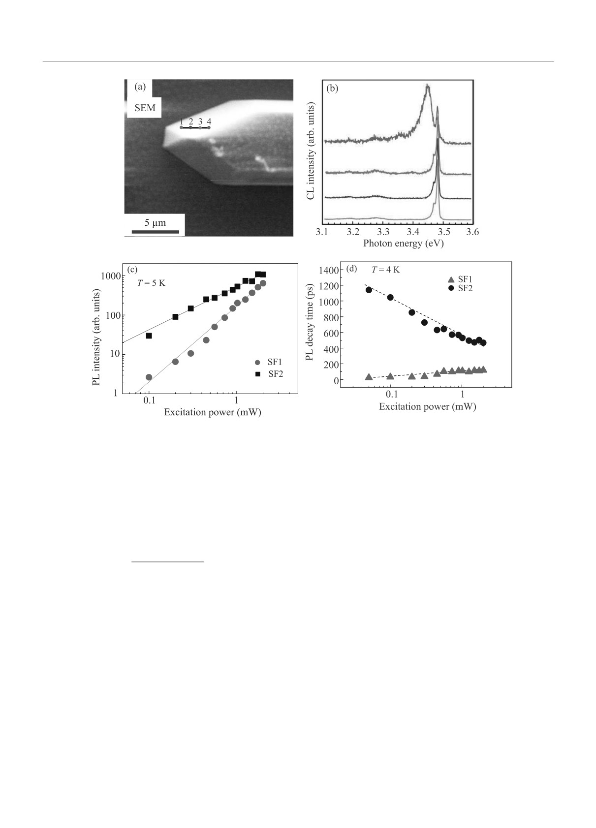
An increase in the excitation power P leads to a
nar nanostructures attract significant interest due to a
super-linear growth of the integrated intensity I ∼ P1.9
relative simplicity of growth [1-4].
for the SF1 emission compared to the SF2 line, for which
We have studied microcavities based on GaN nanos-
the dependence of the integrated PL intensity shows
tructures grown using a “bottom-up” approach [5, 6].
nearly linear growth on pumping I ∼ P1.1 (Fig. 1с).
Near the perfect shape of structures grown by selective
Studies of time-resolved PL have revealed that the
area metal-organic vapor phase epitaxy (MOVPE) on
SF1 and SF2 emissions have opposite temporal behav-
sapphire (0 0 0 1) allowed the formation of cavity modes
ior with the variation of the excitation power. The es-
and the enhancement of the spontaneous emission in-
timated PL decay time is plotted as a function of the
tensity [7, 8]. In such microcavities, the exciton is con-
pumping power in Fig. 1d for the PL lines SF1 and SF2
sidered as “bulk”, (not confined in the active layer in
by solid triangles and circles, respectively. Clearly, the
difference from the microcavities with DBR). Exciton-
recombination time is shorter for the SF1 emission com-
cavity modes coupling conditions are different for these
pared to the SF2 line. As the excitation power increases,
resonators.
τ increases for the SF1 line from ∼ 30 to ∼ 100 ps while,
We compare microcavities formed by GaN nanowires
in contrast, τ decreases from ∼1100 to ∼ 500 ps for the
with structural defects (basal-plane stacking faults
SF2 emission.
(SFs)) and without them and observe different behavior
Interaction of exciton and cavity modes can occur in
of the PL intensity and PL decay time with increasing
microcavities of various types; the resonator size should
excitation power for the exciton localized at SFs com-
be significantly smaller than the wavelength to pro-
pared to the bulk exciton.
vide a large energy interval between the microcavity
Figure 1a shows the end of a uniform flat GaN
modes [10]. The studied planar GaN NWs have width
nanowire. The structures may have structural defects
and length of ∼ 7 and ∼ 110 µm, respectively, and, thus,
such as SFs. The low-temperature CL spectra shown in
possess properties of so-called meso-cavity when the
Figs. 1b were taken at different points of the nanowire
size of the resonator corresponds to tens of wavelengths
along the line as shown in Fig. 1a. Panchromatic CL
[11, 12]. The field distribution for the cavity modes was
shows non-uniformity in the contrast at the edge of
calculated. It is shown that light is well localized at the
the NW, which indicates a presence of structural de-
ends of nanowires.
fect likely SFs. While CL spectra in points 1-3 in
Fig. 1b shows near band gap emission with one domi-
In conclusion, a different behavior of the PL inten-
nant peak at ∼ 3.48 eV, which is typical donor bound
sity and the PL decay time with increasing excitation
exciton (DBE) emission in GaN, the CL spectrum in
power for an exciton localized on a SF as compared to
point 4 demonstrates additional broader emissions at
a bulk exciton is demonstrated. Calculations show good
∼ 3.43 and ∼ 3.35 eV. Similar signatures at ∼ 3.42 eV
localization of the field at the boundary of the structure
and ∼ 3.35 eV in GaN are associated with the emissions
in the region of the exciton energy.
related to the basal plane SFs of type I1 and I2 [9,10].
The work has been supported by the Russian Sci-
ence Foundation # 21-12-00304. The work is financially
1)e-mail: ilinishna@gmail.com
supported by Priority 2030 program.
Письма в ЖЭТФ том 115 вып. 9 - 10
2022
611
5∗