

Журнал эволюционной биохимии и физиологии, 2020, T. 56, № 7, стр. 758-758
Ammocoete – Lamprey Larva (Lampetra planeri Bloch.) Systemic Heart Sinus Venous Cells Are Tolerant to Hypocalcemia
V. A. Golovko 1, *, A. V. Kozlovskaya 2
1 Institute of Physiology, Federal Science Centre KomiSC, Ural Branch RAS
Syktyvkar, Russia
2 Medical Institute of Federal State Syktyvkar University
Syktyvkar, Russia
* E-mail: golovko@physiol.komisc.ru
It is known that changing the solution “without CaCl2” to normal ones causes irreversible damage in the myocardium sarcolemma and intracellular structures due to overload with Ca and Na ions. The aim of our work was to investigate the effects of Ca2+ cycling, and ryanodine and nifedipine on electrical activity of the ammocoete sinus venous cells. The study was performed with using the microelectrode leads technique on the spontaneously shrinking strips of 2 × 3 (mm) of sino-atrial (SA) region. Larvae were anesthetized in 0.1% zaletil. The strips generated triangular-shaped Alice potentials with spike action potential duration of about 0.1 s spontaneously. Slow diastolic depolarization (SDD) was 80–90% of the cycle duration. The minimum speed of SDD and AP generation frequency of the ammocoete sinus venous cells (17–25 imp/min) were observed at the concentration of calcium in the solution from 0.9 to 1.8 mmol (Fig. 1). The highest frequency of AP (98 imp/min), was in solution "without Ca”. The decrease in the frequency of AP generation by 10–15% was registered in solution “without Ca” in the first 10–15 min, and then the frequency increased by 20–30% relative to the control (n = 7; p < 0.05). After 1 hour, a decrease in the pulse frequency was observed again. The strips continued to generate AP and contraction slightly for 6 hours or more. In calcium depletion, AP amplitude decreased from 91 to 75 mV in due to overshoot decrease. The decrease in AP50 duration by 20% was registered also. Similar effects were observed after exposure of nifedipine (2 µm) or ryanodine (20 µm). Thus, it was found that the effect of hypocalcemia in the cells of the ammocoete’s pacemaker is completely reversible after reperfusion. Probably, the cells of the venous sinus of the ammocoete are able to regulate the intake of Ca ions and generate AP in solution “without Ca” due to the inward current INa and the outward IKr. The use of an evolutionary approach allows us to deepen the understanding of the mechanisms of the pacemaker activity formation.
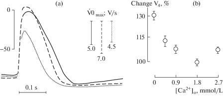
Fig. 1.
Effects of variation of [Ca2+]o 0.9 (continuous line), 0.45 mmol (dashed line) and salt solution without addition of calcium (dotted line) on APs configuration and the upstroke velocity (dV/dt max) (a) and the rate of diastolic depolarization (b). APs traces were recorded in the same cell as in (a).
Supported by SRW Projects GR (AAAA-A17-117012310152-2, AAAA-A17-117012310154-6).
Список литературы отсутствует.
Дополнительные материалы отсутствуют.
Инструменты
Журнал эволюционной биохимии и физиологии