

Журнал эволюционной биохимии и физиологии, 2020, T. 56, № 7, стр. 803-803
The Fusion of Nerve Processes and the Formation of Syncytia in Living Mollusk Neurons in Tissue Culture
O. S. Sotnikov *
Pavlov Institute of Physiology RAS
Saint Petersburg, Russia
* E-mail: ossotnikov@mail.ru
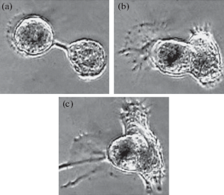
The function of interneuronal gap junctions is considered the main way of electrical interconnection of neurons and an important reticular complement to neural theory. However, there is a lot of experimental data indicating additional possibilities of synсytial connections in the nervous system rather than synapses. The first specific morphological data on the cytoplasmic fusion of nerve processes were identified in polychaetes by a convinced neuronist (Retzius, 1884), and in humans by a reticularist A. S. Dogiel (1893). However, syncytium, as a common structure of many cell types, is almost unknown in neurology.A search was made for all the different forms of interneuronal syncytium in living mollusk neurons in tissue culture (Fig. 1 Syncytial commissure between two neurons and their cytoplasmic fusion. Culture of neurons. Phase contrast. Vol. 40Ph, approx. 10). The most convincing result was fusion of end-to-end fibers of two closely spaced neurons. Most of these fused, stable or having contractions processes, should be considered normal electrical syncytial anastomoses. The fusion of regenerating processes end-to-end usually forms a ring-shaped fiber syncytium. Growing new growth cones, fusing, form two-story syncytial connections. If the branches of the next two branches are located next to each other, they can touch and fuse sides. Branches of several neurons, forming a bundle, fuse into a single nerve fiber. Parts of the bend of the process, in contact with the neighboring one, also form a syncytial contact. Syncytium formation begins with the formation of a gap junction and ends with the breakthrough of two contacting membranes and sealing of the edges of the perforation. The appearance of syncytial fusion of a large number of thin branches forms a network that forms a reticular fragment of nerve tissue.
Список литературы отсутствует.
Дополнительные материалы отсутствуют.
Инструменты
Журнал эволюционной биохимии и физиологии