

Микология и фитопатология, 2023, T. 57, № 2, стр. 141-143
Micromycetes Rossicae: Chorological and Taxonomical Notes. 5. Pseudocercosporella Filipendulae (Mycosphaerellales, Ascomycota) – New Find for Saint Petersburg (Russia)
1 Komarov Botanical Institute of the Russian Academy of Sciences
197376 St. Petersburg, Russia
* E-mail: iv_zmitrovich@mail.ru
Поступила в редакцию 15.11.2022
После доработки 22.12.2022
Принята к публикации 28.12.2022
- EDN: NJJYWJ
- DOI: 10.31857/S0026364823020137
Аннотация
The present report continues the series devoted to rare and interesting species of microfungi from various regions of Russia that cause rust, shoot deformations or leaf spots, and highlights Pseudocercosporella filipendulae (Mycosphaerellales, Ascomycota), a rare species new to Saint Petersburg City (Russia). The morphology of the species was repeatedly studied, emphasizing the conidia variability. This note gives further information on the distribution of the species in Russia, as well as the size of its conidia.
The present report continues a series devoted to rare and interesting species of microfungi of various regions of Russia that cause rust, shoot deformations or leaf spots (Zmitrovich et al., 2020a, 2020b; Dudka, Zmitrovich, 2020, 2021) and highlights a less known representative of the genus Pseudocercosporella.
The genus Pseudocercosporella was described by Deighton (1973) for phytopathogenic, leaf spots-forming cercosporoid fungi with colorless/pale hyphae, well-developed to reduced (mostly hyaline) stromata, semi-micronematous to macronematous, simple, rarely branched, straight and subcylindric to geniculate-sinuous, continuous (reduced to a single conidiogenous cell) or septate, solitary to fasciculate conidiophores forming flat, crustose to subglobose sporodochial conidiomata, separate or integrated, terminal, monoblastic, determinate to polyblastic, sympodial, indeterminate conidiogenous cells (conidiophores reduced to a single conidiogenous cell), and inconspicuous, thin-walled, hyaline, not darkened, subcylindrical, filiform or somewhat obclavate, euseptate, usually multiseptate, conidia of schizolytic separation pattern.
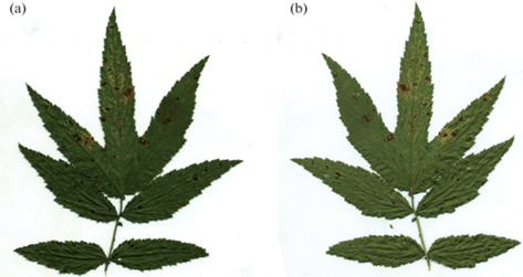
Fig. 1.
Symptoms caused by Pseudocercosporella filipendulae on Filipendula ulmaria (LE 287681): а – the upper leaf surface; b – the lower leaf surface. Scale bar – 1 cm.
In light of molecular data, the genus appears to be polyphyletic (Frank et al., 2010), and not every species described so far have been verified molecularly. A number of species are known from scarce finds. In this notice, we focus on Pseudocercosporella filipendulae, previously known from two finds in Russia.
This taxon was originally described as Cercospora laxipes f. filipendulae by Mel’nik (1966) based on some differences from f. laxipes in conidia size and substrate specialization (“Ad forma laxipes sporulis minutibus (28–60 × 3–4 µm) et hospitis Filipendula ulmaria (L.) Maxim differ”, ibid.). The type specimen was collected in the Russian Soviet Federative Socialist Republic (R.S.F.S.R.), Leningrad Region (Tosnensky district, near Lisino-Korpus village), in a swamp spruce forest of, on fresh leaves of Filipendula ulmaria. Later, Braun and Mel’nik (2008) raised the rank of this taxon to species and placed it in the genus Pseudocercosporella, based on the morphological concept of the genus. Also, these authors have presented the second find of Pseudocercosporella filipendulae from the Pskov region of Russia (Pechersky district, near Livimae station, also on living leaves of Filipendula ulmaria). We found this rare species in September 2022 in Saint Petersburg City (Pushkin, Babolovsky Park) on the same host. This is its third find in Russia; the new material collected broadens the understanding of the range of variabi-lity of the species’ microstructures.
Macroscopic photographs were done using a Nikon D80 camera with AF Micro Nikkor 60 mm lens. Micromorphological analysis of lesions was performed using a Zeiss AxioImager.A1 light microscope. Micropreparations were mounted in distilled water or a 5% KOH solution. The sizes of conidiophores and conidia were measured in 40–60 times random replicates in water. The variability of conidia was assessed according to the methods proposed by Parmasto et al. (1987).
Pseudocercosporella filipendulae (Melnik) U. Braun et Melnik, Mikol. Fitopatol. 42 (4): 305, 2008. – Cercospora laxipes J.J. Davis f. filipendulae Melnik, Novit. Syst. Pl. non Vasc. 3: 218, 1966.
Description. Colonies hypophyllous, effuse, arachnoid or patches-forming, whitish-gray. Mycelium both internal and external. Vegetative hyphae septate, branched, hyaline, smooth, varies in diameter as:
Stromata absent or reduced to slightly swollen basal cells. Conidiophores arising on superficial or substomatal mycelium, solitary or loose fasciculate, unbranched, more or less erect, straight or geniculate, cylindrical, 0–1-septate, with 1–3 conidiogenous loci. Conidia solitary, obclavate to hyphoid or filifom, sometimes sublunate, hyaline, thin-walled, smooth, (0) 1–10-septate, varying in their sizes as:
| Mel’nik (1966) | Braun, Mel’nik (2008) | current data | |
| length × diameter in medial part | 28–60 × 3–4 μm | (10) 25–70 (85) μm | (14) 22–60 (82) μm |
| diameter at the base | 1–2 μm2 | 1–1.5 μm | (0.5) 1–1.5 (2.0) μm |
Symptoms. Leaf spots amphigenous, necrotic, diffuse to angular, 0.2–1.5 mm in diam., sometimes confluent in clusters up to 4 mm in diam., on the upper leaf surface first rose-red, then cinnamon-brown, on the lower leaf surface cinnamon-brown, sometimes with yellowish areolation.
Hosts. Only one host is known so far, namely Filipendula ulmaria (Rosaceae).
Distribution range. Russia: Leningrad Region, Pskov Region, Saint Petersburg.
Material examined: R.S.F.S.R., Leningrad Region, Tosno Rayon, Lisino-Korpus, 08.08.1963, on F. ulma-ria, leg. et det. V.A. Mel’nik (LE 40409, holotype of Cercospora laxipes f. filipendulae). – Russia, Pskov Region, Pechery Rayon, Livimae, 11.08.2007, on F. ulmaria, leg. G.Yu. Konechnaya, det. V.A. Mel’nik (LE 232213). – Russia, Saint Petersburg, Pushkin, Babolovsky Park, 25.09.2022, on F. ulmaria, leg. et det. I.V. Zmitrovich (LE 287681).1122
In other countries, according to our data, this species has not been recorded till now. It is possible that, like all members of the genus, this species is host-specific. North American Pseudocercosporella crataegi is morphologically close to P. filipendulae, however, is distinguished by the regular formation of well-deve-loped conidiophore fascicles, emerging through stomata, as well as larger conidia, reaching 120 × 5 µm and having up to 12 septa (Braun, 1995).
For all known localities, the host plant Filipendula ulmaria is a species of native flora. It is a species gravitating toward more or less aerated wetlands on black woody peat. In Saint Petersburg, the habitat is located in the undrained part of a park with vegetation in its natural dynamic phase (birch-alder swamp forest). All known finds of the pathogen are located in the southern taiga subzone, no material has been yet obtained from the middle taiga subzone.
The work was carried out using technique of the Center “Cellular and Molecular Technologies for Studying Plants and Fungi” at Komarov Botanical Institute of the Russian Academy of Sciences and was supported by the State Research Task (BIN RAS, № 122011900033-4).
Список литературы
Braun U. A monograph of Cercosporella, Ramularia, and allied genera (phytopathogenic Hyphomycetes). IHW-Verlag, Eching, 1995.
Braun U., Mel’nik V.A. Pseudocercosporella filipendulae – a new hyphomycete species from Russia. Mikologiya i fitopatologiya. 2008. V. 42 (4). P. 305–307.
Deighton F.C. Studies on Cercospora and allied genera. IV. Cercosporella Sacc., Pseudocercosporella gen. nov. and Pseudocercosporidium gen. nov. Mycol. Papers. 1973. V. 133. P. 1–62.
Dudka V.A., Zmitrovich I.V. Micromycetes Rossicae: Chorological and taxonomical notes. 3. Exobasidium sundstroemii (Exobasidiales, Basidiomycota) – new find for Leningrad Region. Mikologiya i fitopatologiya. 2020. T. 54, № 6. P. 460–464. https://doi.org/10.31857/S0026364820060069
Dudka V.A., Zmitrovich I.V. Micromycetes Rossicae: chorological and taxonomical notes. 4. Sphacelotheca hydropiperis and Microbotryum cordae (Pucciniomycotina, Microbotryomycetes), two difficult to detection Persicaria-associated micromycetes, new for Volgograd Region (Russia) // Mikologiya i fitopatologiya. 2021. T. 55. № 6. C. 453–456. https://doi.org/10.31857/S0026364821060076
Frank J., Crous P.W., Groenewald J.Z. et al. Microcyclospora and Microcyclosporella: novel genera accommodating epiphytic fungi causing sooty blotch on apple. Persoonia. 2010. V. 24. P. 93–105. https://doi.org/10.3767/003158510X510560
Mel’nik V.A. New imperfect fungi. I. Novosti sistematiki nizschikh rasteniy. 1966. V. 3. P. 214–218 (in Russ.).
Parmasto E., Parmasto I., Möls T. Variation of basidiospores in the Hymenomycetes and its significance to their taxo-nomy. Bibltheca Mycol. 1987. V. 115. P. 1–168.
Zmitrovich I.V., Dudka V.A., Shevchuk S.V. Micromycetes Rossicae: Chorological and taxonomical notes. 1. Chrysomyxa succinea (Pucciniales, Basidiomycota) – new find for Saint Petersburg, European Russia. Mikologiya i fitopatologiya. 2020. V. 54 (4). P. 305–308. https://doi.org/10.31857/S0026364820040133
Zmitrovich I.V., Dudka V.A. Micromycetes Rossicae: Chorological and Taxonomical notes. 2. Melampsora arctica (Pucciniales, Basidiomycota) – urediniospore variability in specimens from European and Siberian Arctic. Mikologiya i fitopatologiya. 2020. V. 54 (5). P. 384–388. https://doi.org/10.31857/S0026364820050128
Мельник В.А. (Mel’nik) Новые несовершенные грибы. I // Новости систематики низших растений. 1966. Т. 3. С. 214–218.
Дополнительные материалы отсутствуют.
Инструменты
Микология и фитопатология