

Зоологический журнал, 2019, T. 98, № 3, стр. 278-284
Contribution to the knowledge of the oribatid mite genus Similobates Mahunka 1982 (Acari, Oribatida, Scheloribatidae)
1 Тюменский государственный университет
625003 Тюмень, Россия
* E-mail: ermilovacari@yandex.ru
Поступила в редакцию 25.01.2018
После доработки 24.06.2018
Принята к публикации 23.05.2018
Аннотация
A new species of Similobates (Oribatida, Scheloribatidae) is described from Cameroon. Similobates staryi sp. n. differs from S. deficiens (Wallwork 1977) and S. demetororum Mahunka 1982 in distinctive morphological traits, e.g., body surface, structure of rostrum, presence of translamellar line, structure and length of bothridial setae, localization of bothridia, as well as lengths of notogastral setae p1 and adanal setae ad1 and ad2. A revised generic diagnosis of, an identification key to, and data on the distribution and ecology of the representatives of Similobates are provided.
The oribatid mite genus Similobates (Acari, Oribatida, Scheloribatidae) was proposed by Mahunka (1982) with Similobates demetororum Mahunka 1982 as type species. At present, the genus comprises two species, which are distributed in the Ethiopian region (Mahunka, 1982; Wallwork, 1977).
Among the material collected from Korup National Park of Cameroon, I found one new species of Similobates. The main goals of the paper are: (a) to describe and illustrate a new species, (b) to propose a revised generic diagnosis, (c) to provide an identification key to known taxa of Similobates, and (d) to present information on the distribution and ecology of the representatives of the genus.
METHODS
Specimens were mounted in lactic acid on temporary cavity slides for measurement and illustration. Body length was measured in lateral view, from the tip of the rostrum to the posterior edge of the notogaster. Notogastral width refers to the maximum in dorsal aspect behind pteromorphs. Lengths of body setae were measured in lateral aspect. All body measurements are presented in micrometers. Formulas for leg setation are given in parentheses according to the sequence trochanter–femur–genu–tibia–tarsus (famulus included). Formulas for leg solenidia are given in square brackets according to the sequence genu–tibia–tarsus. Morphological terminology used in this paper follows that of F. Grandjean: see Travé and Vachon (1975) for references, Norton (1977) for leg setal nomenclature, and Norton and Behan–Pelletier (2009), for overview. Drawings were made with a camera lucida using a Leica transmission light microscope “Leica DM 2500”.
The following abbreviations are used on the figures: ro, le, in, bs, ex – rostral, lamellar, interlamellar, bothridial and exobothridial setae, respectively; kf – keel-shaped ridge; t – tooth; Al – sublamellar porose area; D – dorsophragma; P – pleurophragma; Sa, S1, S2, S3 – notogastral saccules; c, la, lm, lp, h, p – notogastral setae; ia, im, ip, ih, ips – notogastral lyrifissures; Amar – marginal porose area; gla – opisthonotal gland opening; h, m, a – subcapitular setae; or – adoral seta; v, l, d, cm, acm, ul, sul, vt, lt – palp setae; ep – postpalpal seta; cha, chb – cheliceral setae; Tg – Trägårdh’s organ; Pd I, Pd II – pedotecta I, II, respectively; 1a, 1b, 1c, 2a, 3a, 3b, 3c, 4a, 4b, 4c – epimeral setae; dis – discidium; cp – circumpedal carina; g, ag, an, ad – genital, aggenital, anal and adanal setae, respectively; p.o. – preanal organ; f – furrow; iad – adanal lyrifissure; ω, σ, φ – solenidia; ɛ – famulus; p.a. – porose area; pr – process; Tr, Fe, Ge, Ti, Ta – trochanter, femur, genu, tibia, tarsus, respectively.
The following abbreviations of collections are used: SMNH – Senckenberg Museum of Natural History, Görlitz, Germany; TSUMZ – Tyumen State University Museum of Zoology, Tyumen, Russia.
SYSTEMATICS
Genus Similobates Mahunka 1982
Type species: Similobates demetororum Mahunka 1982
Revised generic diagnosis
Adult. Body surface without heavy sculpture and ornamentation, sometimes partially foveolate. Rostrum rounded or pointed. Lamellae well-developed, thin. Translamella absent or represented by short parts near lamellae. Sublamellae, sublamellar porose areas and lateral keel-shaped ridges present. Prolamellae and tutoria absent. Pteromorphs broad, rounded. Prodorsal setae long, setiform; insertions of rostral setae clearly distanced. Bothridial setae short or long, setiform, lanceolate or globular. Notogaster oval, slightly longer than wide. Anterior notogastral margin convex, covering or not bothridial openings. Dorsophragmata semioval or elongated. Notogaster with four pairs of small saccules and 10 pairs of minute setae, rarely only p1 long. Epimeral setal formula 3–1–3–3. Pedotecta II rounded or bifurcate distally. Five pairs of genital, one pair of aggenital, two pairs of anal and three pairs of adanal setae. Adanal setae of medium size or long, ad3 inserted in preanal position. Adanal lyrifissures located in paraanal position, close and parallel to anal plates. Leg tarsi with three claws.
Juvenile instars. Not known.
Remarks. The genus Similobates differs mostly from a morphologically similar Scheloribates Berlese 1908 by the number of genital setae (five pairs versus four). Although I presently support the initial generic status of Similobates, additional research is necessary for a better understanding of its status.
Similobates staryi Ermilov sp. n. (Figs 1–3)
Fig. 1.
Similobates staryi sp. n., adult: a – dorsal view (legs not shown); b – ventral view (gnathosoma and legs not shown); c – subcapitulum, ventral view; d – palp, right, antiaxial view; e – chelicera, left, paraxial view. Scale bar (µm): a, b – 100; c–e – 50.
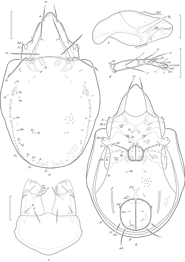
Fig. 2.
Similobates staryi sp. n., adult: a – anterior part of body, lateral view (gnathosoma and legs not shown); b – posterior part of body, lateral view; c – posterior view. Scale bar 100 µm.
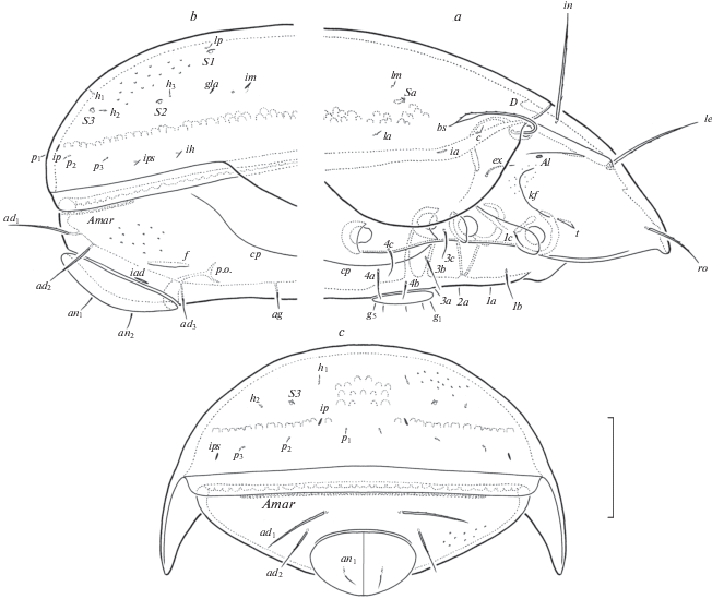
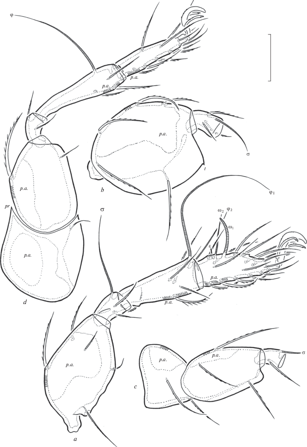
Fig. 3.
Similobates staryi sp. n., adult: a – leg I, without trochanter, right, antiaxial view; b – femur and genu of leg II, right, antiaxial view; c – trochanter, femur and genu of leg III, left, antiaxial view; d – leg IV, left, antiaxial view. Scale bar 50 µm.
Material. Holotype (♂) and 16 paratypes (8 ♀♀, 8♂♂): Cameroon, South-West Province, Korup National Park, Rengo Camp, about 8 km NW of Mundemba, latitude 05°02′11.64″ N, longitude 08°49′45.96″ E, altitude 300 m, litter and soil sifting sample, 12–16.V.2006 (V.V. Grebennikov).
The holotype (ethanol with drop of glycerol) and two paratypes (ethanol with drop of glycerol) are deposited in SMNH; 14 paratypes (ethanol with drop of glycerol) are deposited in TSUMZ.
Diagnosis. Body size: 481–780 × 265–448. Notogaster and anogenital region foveolate. Rostrum narrowly rounded. Translamellar line represented by two rudimentary parts near lamellae. Prodorsal setae setiform, barbed, ro shortest, in longest. Bothridial setae long, lanceolate, barbed. Lateral parts of prodorsum with one pair of teeth. Notogastral setae minute. Epimeral setae setiform, slightly barbed. Circumpedal carinae long. Genital, aggenital, anal and adanal setae ad3 setiform, thin, indistinctly barbed, adanal setae ad1 and ad2 thicker, erect, barbed. Marginal porose area band-like. Leg tarsi I and II with one or two teeth dorsally, femora II with tooth ventroanteriorly, trochanters IV with process anterodorsally. Leg tarsi I with 19 setae (l '' absent).
Description. Measurements. Body length: 481 (holotype, male), 481–780 (paratypes); notogaster width: 265 (holotype), 265–448 (paratypes). No clear differences between females and males in body size, but females usually larger.
Integument (Figs 1a, 1b; 2a; 3b, 3c). Body color light brown to brown. Body surface and legs microfoveolate (visible under high magnification). Lateral parts of prodorsum between sublamellae and acetabula I, II microgranulate. Notogaster and anogenital region sparsely foveolate (diameter of foveoles up to 4).
Prodorsum (Figs 1a; 2a). Rostrum slightly protruding, narrowly rounded. Lamellae located dorsolaterally, about half of prodorsum (measured in lateral view). Sublamellae thin, about 2/3 of lamellae. Translamellar line represented by two short, rudimentary parts near lamellae. Sublamellar porose areas oval (6–10 × × 4–8). Rostral (57–61 up to 73 in some paratypes), lamellar (77–90 up to 106 in some paratypes) and interlamellar (110–118 up to 131 in some paratypes) setae setiform, sparsely barbed, in indistinctly thicker than others. Bothridial setae (123–135 up to 164 in some paratypes) lanceolate, sparsely barbed, with long stalk and short head having well visible apex. Anterocentral transverse ridge (between rostral and lamellar setae) and sejugal porose areas not visible. Lateral keel-shaped ridges well-developed. One pair of lateral teeth present anteriorly to kf. Exobothridial setae (32–41) setiform, slightly barbed. Dorsophragmata elongated.
Notogaster (Figs 1a; 2a–2c). Anterior notogastral margin convex medially. Ten pairs of notogastral setae minute (4). Four pairs of saccules with small openings and drop-like channels. Distance S1–S1 shorter than S2–S2. Setae lm inserted posteromedial to Sa, lp medial to S1. All lyrifissures distinct; im located between Sa and h3, ip between p1 and S3, ih and ips distanced from each other. Opisthonotal gland openings located posterior to im. Circumgastric scissure and circumgastric sigillar band distinct.
Gnathosoma (Figs 1c–1e). Subcapitulum longer than wide (155–159 × 110–123). Subcapitular setae setiform, slightly barbed; h (30–32) longer than a (26–28) and m (24–26), a thickest, m thinnest. Two pairs of adoral setae (18–20) setiform, heavily barbed. Palps (length 98–102) with typical setation 0–2–1–3–9(+ω). Postpalpal setae (6) spiniform, smooth. Chelicerae (length 172–176) with two setiform, barbed setae, cha (53–57) longer than chb (36–41). Trägårdh’s organ of chelicerae elongate triangular.
Epimeral and lateral podosomal regions (Figs 1b; 2a). Epimeral setal formula: 3–1–3–3. Setae setiform, thin, slightly barbed; 1b, 3b, 3c, 4a, 4c (24–28) longer than 1c, 4b (16–20) and 1a, 2a, 3a (12–16). Setae 1c inserted laterally on pedotecta I. Pedotecta I and II represented by small laminae, Pd II bifurcate in ventral view. Discidia slightly developed, rounded distally. Circumpedal carinae long, directed to pedotecta II.
Anogenital region (Figs 1b; 2a–2c). Five pairs of genital (g1, 16–20; g2–g5, 12–16), one pair of aggenital (16–20), two pairs of anal (16–20) and one pair of adanal (ad3, 16–20) setiform, thin, indistinctly barbed. Two pairs of adanal setae (ad1, 57–61 up to 73 in some paratypes; ad2, 36–41 up to 53 in some paratypes) setae setiform, erect, sparsely barbed. Adanal lyrifissures located close and parallel to anal plates. Adanal setae ad1 in posterior, ad2 in posterolateral, ad3 in preanal positions. One pair of longitudinal furrows located anterolateral to anal aperture. Marginal porose area present, complete, band-like. Preanal organ goblet-like. Ovipositor is typical for Scheloribatidae (Ermilov, 2010): elongated (217 × 41), blades (94) shorter than length of distal section (beyond middle fold; 123). Each of the three blades with four setae, ψ1 ≈ τ1 (36) setiform, longer than thorn-like ψ2 ≈ τa ≈ τb ≈ τc (16), all smooth. Six coronal setae spiniform (4).
Legs (Figs 3a–3d). Median claws thicker than laterals, all serrate dorsally. Tarsi I and II with one with one or two small teeth dorsally. Femora II with tooth ventroanteriorly. Trochanters IV with strong process anterodorsally. Dorsoparaxial porose areas on femora I–IV and on trochanters III, IV and ventral porose areas in basal parts of tarsi and distal parts of tibiae well visible. Formulas of leg setation and solenidia: I (1–5–3–4–19) [1–2–2], II (1–5–3–4–15) [1–1–2], III (2–3–1–3–15) [1–1–0], IV (1–2–2–3–12) [0–1–0]; homology of setae and solenidia indicated in Table 1. Famulus of tarsi I short, erect, slightly dilated and truncated distally, inserted posterior to solenidion ω2.
Table 1.
Leg setation and solenidia of Similobates staryi sp. n.
| Leg | Tr | Fe | Ge | Ti | Ta |
|---|---|---|---|---|---|
| I | v' | d, (l), bv'', v'' | (l), v', σ | (l), (v), φ1, φ2 | (ft), (tc), (it), (p), (u), (a), s, (pv), v', (pl), ɛ, ω1, ω2 |
| II | v' | d, (l), bv'', v'' | (l), v', σ | (l), (v), φ | (ft), (tc), (it), (p), (u), (a), s, (pv), ω1, ω2 |
| III | l ', v' | d, l ', ev' | l ', σ | l ', (v), φ | (ft), (tc), (it), (p), (u), (a), s, (pv) |
| IV | v' | d, ev' | d, l ' | l ', (v), φ | ft'', (tc), (p), (u), (a), s, (pv) |
Remarks. Distinctive characteristics of the new species versus other Similobates species can be found in the identification key below.
Etymology. The species name is dedicated to our colleague and the well-known acarologist Dr. Josef Starý (Biology Centre v.v.i., Czech Academy of Sciences, Institute of Soil Biology, České Budějovice, Czech Republic) for his extensive contributions to our knowledge of oribatid mites.
KEY TO KNOWN SPECIES OF SIMILOBATES
1. Rostrum pointed; bothridial setae globular, very short; bothridia completely covered by anterior margin of notogaster; adanal setae ad1 and ad2 clearly shorter than width of anal plate; body size: 1096–1131 × × 765–817 ….... Similobates deficiens (Wallwork 1977)
– Rostrum rounded; bothridial setae with distinct or indistinct lanceolate head, comparatively long; bothridia not completely covered by anterior margin of notogaster; adanal setae ad1 and ad2 not shorter than width of anal plate …............................................ (2)
2. Notogaster and anogenital region foveolate; translamellar line represented by two very short, rudimentary parts near lamellae; bothridial setae with well-developed heads; notogastral setae p1 minute; body size: 481–780 × 265–448 …………. Similobates staryi sp. n.
– Notogaster and anogenital region not foveolate; translamellar line absent; bothridial setae with indistinct heads to setiform; notogastral setae p1 comparatively long; body size: 775–816 × 488–52 …... Similobates demetororum Mahunka 1982
DISTRIBUTION AND ECOLOGY OF SIMILOBATES IN THE WORLD
At present, three species of Similobates (including a new species described in this paper) are distributed in the Ethiopian region, and each species is known from one country only.
Similobates demetororum was recovered from decaying litter in Eucalyptus and Juniperus woodland, near the Akaki River in Addis-Ababa, Ethiopia (Mahunka, 1982). Also, this species was recovered from soil, litter and moss in the Harenna Forest of Bale Mountains National Park, as well as from litter and moss of the Cholomu Forest, located to the North of Ginchi city (Ermilov et al., 2011, 2012; Ermilov, Rybalov, 2013). Similobates deficiens was described from the High Central Ridge of Saint Helena Island, without a biotope designation (Wallwork, 1977). Similobates staryi sp. n. is known from litter and soil of the Korup National Park in Cameroon (see above in the “Material” subsection).
Список литературы
Ermilov S.G., 2010. The structure of ovipositors in higher oribatid mites (Acari, Oribatida, Brachypylina) // Zoologichesky Zhurnal. V. 89. № 6. P. 694–702 (English version: 2010. Entomological Review. V. 90. № 6. P. 783–792).
Ermilov S.G., Rybalov L.B., 2013. Two new species and new records of oribatid mites (Acari: Oribatida) from Ethiopia // Annales Zoologici. V. 63. № 1. P. 45–55.
Ermilov S.G., Rybalov L.B., Franke K., 2011. Ethiopian oribatid mites of the family Scheloribatidae (Acari: Oribatida) // African Invertebrates. V. 52. № 2. P. 311–322.
Ermilov S.G., Sidorchuk E.A., Rybalov L.B., 2012. Oribatid mites (Acari: Oribatida) of Ethiopia // Zootaxa. № 3208. P. 27–40.
Mahunka S., 1982. Oribatids from the Eastern Part of the Ethiopian Region (Acari) I // Acta Zoologica Academiae Scientiarum Hungaricae. V. 28. № 3–4. P. 293–336.
Norton R.A., 1977. A review of F. Grandjean’s system of leg chaetotaxy in the Oribatei (Acari) and its application to the family Damaeidae // Dindal D.L., editor. Biology of oribatid mites. Syracuse, SUNY College of Environmental Science and Forestry. P. 33–61.
Norton R.A., Behan-Pelletier V.M., 2009. Oribatida // A Manual of Acarology (TX). Lubbock: Texas Tech University Press. P. 430–564.
Travé J., Vachon M., 1975. François Grandjean. 1882–1975 (Notice biographique et bibliographique) // Acarologia. V. 17. № 1. P. 1–19.
Wallwork J.A., 1977. 4. Acarina. 4.1. Cryptostigmata // La faune terrestre de L’île de Sainte-Hélène (quatriѐme partie). Tervuren, Musee Royal de L’Afrique Centrale, Belgique Annales. P. 189–257.
Дополнительные материалы отсутствуют.
Инструменты
Зоологический журнал