

Микология и фитопатология, 2020, T. 54, № 6, стр. 460-464
Micromycetes Rossicae: Сhorological and Taxonomical Notes. 3. Exobasidium Sundstroemii (Exobasidiales, Basidiomycota) – New Find for Leningrad Region
V. A. Dudka 1, *, I. V. Zmitrovich 1, **
1 Komarov Botanical Institute of the Russian Academy of Sciences
197376 St. Petersburg, Russia
* E-mail: dudkavasiliy.a@gmail.com
** E-mail: iv_zmitrovich@mail.ru
Поступила в редакцию 1.03.2020
После доработки 1.05.2020
Принята к публикации 11.05.2020
Аннотация
Exobasidium representatives cause various deformations of the aerial part of ericaceous, theaceous, and lauraceous plants. In the course of the present research carried out on the territory of the oceanic sector of the European middle taiga subzone in the vicinity of the Vaskelovo settlement (Leningrad Region), we found rather little-known species, Exobasidium sundstroemii. The morphological features of this species and symptoms demonstrated by infected plants are considered and compared with those of widespread species E. karstenii, more frequent on Andromeda polifolia leaves.
The present notice continues a series devoted to rare and interesting species of micromycetes of various regions of Russia that cause rust and other leaf spots (Zmitrovich et al., 2020a, 2020b).
The Exobasidium species (Basidiomycota, Ustilaginomycotina, Exobasidiomycetes, Exobasidiales) compose a specific parasitic group which provokes the plant to over-production of growth hormones and causes hypertrophy of the affected areas of host tissues. At the current moment, there are about 120 species of the genus found in various climatic zones. The genus representatives affect predominantly the members of the Ericaceae family, but were found also on the members of Theaceae, Lauraceae, and a number of other families. The main symptoms of damage caused by Exobasidium spp., are localized in different parts of the plant, for example, the pathogen forms local spots on host leaves, sometimes causes its thickening, colonizes a young shoot (which leads to the formation of “witch’s brooms”), buds, fruits or form systemic damage, when the leaves of the plant increase in size (hypertrophy and hyperplasia), and sometimes the entire plant becomes entirely red (Nannfeldt, 1981; Karatygin, 2002; Klenke, Scholler, 2015).
The genus Exobasidium is fairly well studied in the European part of the temperate zone. However, despite this, a number of species remain (E. arescens Nannf., E. ledi P. Karst., E. oxycocci Rostr. ex Shear, E. sundstroemii Nannf.) which are rare in the European part of Russia or have a dubious taxonomic status. For example, Döring (2003), using molecular methods, could not confirm the distinction between E. juelianum Nannf. and E. vaccinii (Fuckel) Woronin.
In the present notice, we would like to report about the first record of E. sundstroemii in the Leningrad Region as well as consider its morphological differences from another species, E. karstenii Sacc. et Trotter, more usual on Andromeda polifolia (Ericaceae).
The exobasidiaceous parasite on Andromeda polifolia causing a systemic disease called “rose blossom” for a long time was considered as Exobasidium andromedae Peck. Studying this species in detail, i.e. using morphological, cultural, and physiological approaches, Sundstrӧm (1964) came to the conclusion that this species is not homogeneous, however this researcher not complete this important work. This research was continued by another Norwegian researcher Nannfeldt (1981), who divided E. andromedae into two species, E. karstenii and E. sundstroemii. These works convincingly confirmed the independent status of both species. In recent works, devoted to the phylogeny of the genus Exobasidium, where molecular methods were used, the phylogenetic trees were shown, generated on the basis of nr5.8S, nrSSU, and nrSSU data. These results show that E. sundstroemii and E. karstenii place different position in molecular phylograms (Begerow et al., 2002; Döring, 2003; Piątek et al., 2012; Brewer et al., 2014), therefore, two rather distant Exobasidium phylospecies were adapted to parasitizing the same Andromeda host.
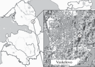
During mycological studies within swamp forests and raised bogs in July 2019, in the vicinity of the Vaskelovo settlement (Leningrad Region), we found E. sundstroemii (Fig. 1). Earlier for Russia, this species was already noted by Gomzhina and Tobias (2015) on Sredniy Isle of the Kretsk Archipelago. The GBIF website also contains a record on E. sundstroemii by Filippova from the Khanty-Mansi Autonomous Okrug of the Tyumen Region (Filippova, 2020). In the present notice, a detailed diagnosis of this species is given and the differentiating features of E. sundstroemii and E. karstenii are analyzed.
Fig. 1.
Eхobasidium sundstroemii locality (E): Russia, Leningrad Region, Vsevolozhsky district, Vaskelovo settlement vicinity, Oxycocco-Sphagnetum, 60°23′27″N, 30°23′40″E.
Leaves herborization was carried out according to a standard recommendation (Geltman, 1995). Dried shoots and leaves were examined using an MBS-3 binocular stereoscopic microscope. Micromorphological analysis of the affected parts of the plant was carried out using an Axio Scope A1 light microscope in the Laboratory of Systematics and Geography of Fungi of the Komarov Botanical Institute of the Russian Academy of Sciences. The material was prepared manually by thin cutting of the blade. To study the general morphology of microstructures, hyphae were prepared using 5% KOH and Cotton Blue reagent. The collected material kept in the Mycological Herbarium of Komarov Botanical Institute (LE F).
Exobasidium sundstroemii Nannf., Symb. bot. Upsal. 23(no. 2): 59, 1981.
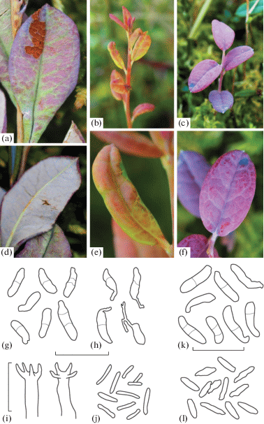
Systemic parasite of Andromeda polifolia. Whole shoot or its parts can be infested. Affected shoots may be crowded. Leaves increase slightly or retain the same size. The edge of the affected leaf is slightly wrapped, the color is pink, red (does not darken over time, as is the case with Exobasidium karstenii), or more often the leaf has a mosaic structure with green patches (Fig. 2, a). A dense white hymenium forms on the leaf abaxial side (Fig. 2d). Basidia cylindrical, usually with 4 (rarely 5) sterigmata (Fig. 2, i). Basidiospores 8.4–14.3 × 1.5–3.7 µm, with a thin wall, hyaline, musiform or ellipsoid, sometimes straightly elongated; there is a distinct chilar appendage. Basidiospores aseptate or with 1 (rarely 3) septa (Fig. 2, g). Basidiospore germination with a seedling tube formation (Fig. 2, h). Conidia 6.7–11 × 0.8–2.0 µm, fusiform, rod-shaped (Fig. 2, j).
Fig. 2.
Symptoms of damage and microstructures of Exobasidium sundstroemii and E. karstenii: a – leaf affected by E. sundstroemii; b, c – plant affected by E. karstenii; d – hymenium of E. sundstroemii on the abaxial side of the leaf; e, f – appearance of diseased leaves of E. karstenii; g – basidiospores of E. sundstroemii; h – germination of E. sundstroemii basidiospores; i – basidia of E. sundstroemii; j – conidia of E. sundstroemii; k – basidiospores of E. karstenii; l – conidia of E. karstenii. Scale bar – 20 μm.
Material examined: Exobasidium sundstroemii, Russia, Leningrad Region, Vsevolozhsky district, Vaskelovo settlement vicinity, Oxycocco-Sphagnetum, 60°23′27″N, 30°23′40″E, on Andromeda polifolia (LE 330119).
General distribution. Russia: Republic of Karelia, Leningrad Region, Tyumen Region (Khanty-Mansi Autonomous Okrug). Europe: Denmark, Estonia, Finland, Germany, Norway, Sweden.
Exobasidium sundstroemii is one of the most hidden and difficult to identify European Exobasidium species. The fact is that it has a rather mild manifestation of lesion symptoms, which often overlap with E. karstenii. As a consequence, this species is often overlooked in field research. Both of these species parasitize the circumpolar Andromeda polifolia. They are characterized by damage to a part of the plant with reddening of the leaves. Moreover, Exobasidium karstenii is characterized by a dark red or almost violet spots production (Fig. 2, c, f). Isolation of the mosaic leaf structure for E. sundstroemii lesions is not confirmed because we found specimens affected by E. karstenii which also caused mosaic damage (Fig. 2, b, e). The number of sterigmata and septa also overlap in both species, therefore, are rather inconstant. These depend on the microclimatic conditions, as pointed out in his work by Gäumann (1922) on the example of closely related genus Kordyana (Exobasidiales). E. sundstroemii (Fig. 2, g, h) is characterized by slightly smaller basidiospores and conidia (Fig. 2, j). E. karstenii, has larger basidiospores (Fig. 2, k), and their conidia are larger and cylindrical (Fig. 2, l).
In our opinion, it is worth paying special attention to two characters when separating these two species, namely, the structure of the mesophyll of affected leaf and the nature of the germination of basidiospores. These characters were identified by Sundstrӧm and Nannfeldt to distinguish between E. sundstroemii and E. karstenii, but for some reason, a number of other researchers (Klenke, Scholler, 2015) omitted these characters from the keys for identification and anamnesis, perhaps to simplify the definition. Table 1 summarizes all the main features of both species with an emphasis on the differential ones.
Table 1.
Differential characteristics of Exobasidium sundstroemii и E. karstenii
| Character | E. sundstroemii | E. karstenii |
|---|---|---|
| Shoots growth | crowded | scattered or less often crowded |
| The nature of shoot affection | partial | systemic, rarely partial |
| Shoots defeat coverage | rarely sprouts from the underground part | often sprouts from underground |
| The nature of the leaf lesion | moderately widened, of normal thickness | greatly enlarged, thin and soft |
| The nature of the defeat of the mesophyll | with typical spongy mesophyll | with undifferentiated mesophyll |
| Leaves color | light purple-red (pink) above, often with green spots | above dark bluish (often almost blackish) purple |
| Hymenium | white on the underside | white on the underside |
| Number of sterigmata | 4–5 | 2–4 |
| Basidiospore sizes | 8.4–14.3 × 1.5–3.7 μm | 11.4–20.0 × 2.0–5.5 μm |
| Basidiospore quotient (Q) | 4.13 | 4.42 |
| Basidiospore shape | musiform, almost straight or moderately geniculate near the hilar appendage | narrowly musiform, moderately allantoid |
| Number of septa in mature basidiospore | 1 (rarely 3) | 1–3 |
| Basidiospore germination | germination by germ-tubes | germination by conidium |
| Conidia | 6.7–11 × 0.8–2.0 μm | 7.4–13.0 × 0.8–2 μm |
| Conidia quotient (Q) | 7.41 | 8.6 |
| Conidia shape | rod-shaped | rod-shaped |
| Frequency | rare | widespread |
Taking into account such ambiguous morphological differences, each affected plant should be carefully examined during parasites identification and all available characters should be recorded. Also, if the identification is difficult, some additional methods can be involved, e.g., molecular data and isolation of the fungus into a pure culture. The mentioned approach is often practiced by Japanese mycologists when studying Exobasidium species on Rhododendron hosts.
The work was carried out using technique of the Center “Cellular and Molecular Technologies for Studying Plants and Fungi” at Komarov Botanical Institute of the Russian Academy of Sciences. Laboratory work was supported by the State Research Task “Herbarium funds of the BIN RAS” (АААА-А18-118022090078-2) and by the RFBR grant (N 19-04-00024 A).
Список литературы
Begerow D., Bauer R., Oberwinkler F. The Exobasidiales: an evolutionary hypothesis. Mycol. Progress. 2002. V. 1. P. 187–199.
Brewer M.T., Turner A.N., Brannen P.M. et al. Exobasidium maculosum, a new species causing leaf and fruit spots on blueberry in the southeastern USA and its relationship with other Exobasidium spp. Parasitic to blueberry and cranberry. Mycologia. 2014. V. 106 (3). P. 415–423. https://doi.org/10.3852/13-202
Döring H. Molekularsystematische Untersuchungen an Vertretern der pflanzenparasitischen Gattung Exobasidium (Basidiomycota). Phd Thesis. Bayreuth, 2003.
Filippova N. The fungarium of Yugra State University reference collection. Version 1.8. Yugra State University Biological Collection (YSU BC). 2020.
Gäumann E. Über die gattung Kordyana Rac. Ann. Mycol. 1922. V. 20. P. 257–271.
Geltman D.V. (ed.). The Herbarium handbook. Kew, 1995 (in Russ.).
Gomzhina M.M., Tobias A.V. Species of Exobasidium Woronin on some islands of Keret Archipelago (White Sea). Vestnik Sankt-Peterburgskogo universiteta. Ser. 3. Biologiya. 2015. N 2. P. 23–31 (in Russ.).
Karatygin I.V. Definitorium fungorum Rossiae. Ordines Taphrinales, Protomycetales, Exobasidiales, Microstromatiales. Nauka, Petropolis, 2002 (in Russ.).
Klenke F., Scholler M. Pflanzenparasitische Kleinpilze. Springer, Berlin, Heidelberg, 2015. https://doi.org/10.1007/978-3-662-46162-4
Nannfeldt J.A. Exobasidium, a taxonomic reassessment applied to the European species. Symb. Bot. Upsal. 1981. V. 23. N 2. P. 1–72.
Piątek M., Lutz M., Welton P. Exobasidium darwinii, a new Hawaiian species infecting endemic Vaccinium reticulatum in Haleakala National Park. Mycol. Progress. 2012. V. 11. P. 361–371. https://doi.org/10.1007/s11557-011-0751-4
Sundstrӧm K.R. Studies of the physiology, morphology and serology of Exobasidium. Symb. Bot. Upsal. 1964. V. 18, N 3. P. 1–89.
Zmitrovich I.V., Dudka V.A., Shevchuk S.V. Micromycetes Rossicae: Chorological and taxonomical notes. 1. Chrysomyxa succinea (Pucciniales, Basidiomycota) – new find for Saint Petersburg, European Russia. Mikologiya i fitopatologiya. 2020. V. 54 (4). P. 305–308. https://doi.org/10.31857/S0026364820040133
Zmitrovich I.V., Dudka V.A. Micromycetes Rossicae: Chorological and Taxonomical notes. 2. Melampsora arctica (Pucciniales, Basidiomycota) – urediniospore variability in specimens from European and Siberian Arctic. Mikologiya i fitopatologiya. 2020. V. 54 (5). P. 384–388. https://doi.org/10.31857/S0026364820050128
Гельтман Д.В. (Geltman) (ред.) Гербарное дело. Справочное руководство. Русское издание. Кью: Королевские ботанические сады, 1995. 342 с.
Гомжина М.М., Тобиас А.В. (Gomzhina, Tobias) Виды рода Exobasidium Woronin на некоторых островах Керетского архипелага (Белое море) // Вестник Санкт-Петербургского университета. Серия 3. Биология. 2015. № 2. С. 23–31.
Каратыгин И.В. (Karatygin) Определитель грибов России. Порядки тафриновые, протомициевые, экзобазидиевые, микростромациевые. СПб.: Наука, 2002. 137 с.
Дополнительные материалы отсутствуют.
Инструменты
Микология и фитопатология