

Зоологический журнал, 2019, T. 98, № 7, стр. 758-772
On Oribatid Mites with Auriculate Pteromorphs in Taiwan (Acari, Oribatida)
S. G. Ermilov *
Tyumen State University
625003 Tyumen, Russia
* E-mail: ermilovacari@yandex.ru
Поступила в редакцию 9.04.2018
После доработки 1.06.2018
Принята к публикации 15.06.2018
Аннотация
Data on the systematics of three species of oribatid mites (Acari, Oribatida) with auriculate pteromorphs from Taiwan are provided. A new species, Neoribates (Neoribates) yangensis sp. n. (Parakalummidae), is described; it is morphologically most similar to Neoribates (N.) similis Fujikawa 2007 and Neoribates (N.) pyramidalis (Tseng 1984), but differs in the body surface structure (lineolate in the new species vs. smooth in N. similis and N. pyramidalis). Descriptive notes on Galumna (Galumna) tsengi Ermilov et Liao 2017 and Galumna (G.) alata (Hermann 1804) (Galumnidae) are also presented.
This work is part of my continuing study of oribatid mites (Acari, Oribatida) from Taiwan (e.g., Ermilov, Liao, 2017, 2017a; Ermilov, 2018), and it includes data on three species with auriculate pteromorphs from the families Parakalummidae (Oripodoidea) and Galumnidae (Galumnoidea).
The primary goal of the paper is to describe and illustrate a new species of Neoribates Berlese 1914 (Parakalummidae). This genus includes four subgenera (see Ermilov, Corpuz-Raros, 2015) and 63 species (Subías, 2004, updated 2018; see also Ermilov, Starý, 2018), which collectively have a cosmopolitan distribution, occurring in all world regions except Antarctica (Subías, 2004, updated 2018). At present, only three species of Neoribates (all from the nominative subgenus) are known from Taiwan: Neoribates (N.) pyramidalis (Tseng 1984), Neoribates (N.) rotundus Aoki 1982 and Neoribates (N.) roubali (Berlese 1910) (Tseng, 1984; Aoki, 1991; Chu, Aoki, 1997).
The secondary goal of the paper is to present a morphological addition to descriptions of Galumna (Galumna) tsengi Ermilov et Liao 2017 and Galumna (G.) alata (Hermann 1804) (Galumnidae) based on specimens from Taiwan.
METHODS
Specimens were mounted in lactic acid on temporary cavity slides for measurement and illustration. Body lengths were measured in lateral view, from the tip of the rostrum to the posterior edge of the notogaster. Notogastral width refers to the maximum width of the notogaster (behind pteromorphs). Lengths of body setae were measured in lateral aspect. All body measurements are presented in micrometers. Formulas for leg setation are given in parentheses according to the sequence trochanter–femur–genu–tibia–tarsus (famulus included). Formulas for leg solenidia are given in square brackets according to the sequence genu–tibia–tarsus.
Drawings were made with a camera lucida using a Leica transmission light microscope “Leica DM 2500”.
Morphological terminology follows that of F. Grandjean (see Ermilov, Klimov, 2017 for review and application).
The following abbreviations are used: lam – lamella; ar, pr, lr – anterior, posterior and lateral ridges of prodorsum, respectively; L – lamellar line; S – sublamellar line; N – prodorsal leg niche; E, T – lateral ridges of prodorsum; ro, le, in, bs – rostral, lamellar, interlamellar and bothridial setae, respectively; Ad – sejugal porose area; D – dorsophragma; P – pleurophragma; c, la, lm, lp, h, p – notogastral setae; Sa, S1, S2, S3 – notogastral saccules; Aa, A1, A2, A3 – notogastral porose areas; mp – median pore; ia, im, ip, ih, ips – notogastral lyrifissures; gla – opisthonotal gland opening; cs – circumgastric scissure; csb – circumgastric sigillar band; h, m, a – subcapitular setae; or – adoral seta; sac – axillary saccule; v, l, d, cm, acm, ul, sul, vt, lt – palp setae; ep – postpalpal seta; ω – palp and leg solenidion; cha, chb – cheliceral setae; Tg – Trägårdh’s organ; Pd I, Pd II – pedotecta I, II, respectively; 1a, 1b, 1c, 2a, 3a, 3b, 3c, 4a, 4b, 4c – epimeral setae; dis – discidium; cp – circumpedal carina; g, ag, an, ad – genital, aggenital, anal and adanal setae, respectively; iad – adanal lyrifissure; Ap – postanal porose area; Amar – marginoventral porose area; p.o. – preanal organ; Tr, Fe, Ge, Ti, Ta – leg trochanter, femur, genu, tibia and tarsus, respectively; p.a. – leg porose area; σ, φ – leg solenidia; ɛ – tarsus I famulus; v, ev, bv, l, d, ft, tc, it, p, u, a, s, pv, pl – leg setae.
The following abbreviations of collections are used: NTU – National Taiwan University, Taipei, Taiwan; TSUMZ – Tyumen State University Museum of Zoology, Tyumen, Russia.
SYSTEMATICS
Neoribates (Neoribates) yangensis Ermilov sp. n. (Figs 1–5)
Fig. 1.
Neoribates (Neoribates) yangensis Ermilov sp. n., adult: dorsal view (legs not shown). Scale bar: 100 μm.
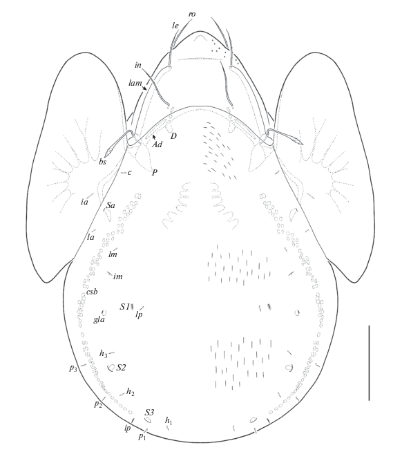
Fig. 2.
Neoribates (Neoribates) yangensis Ermilov sp. n., adult: ventral view (gnathosoma and legs not shown). Scale bar: 100 μm.
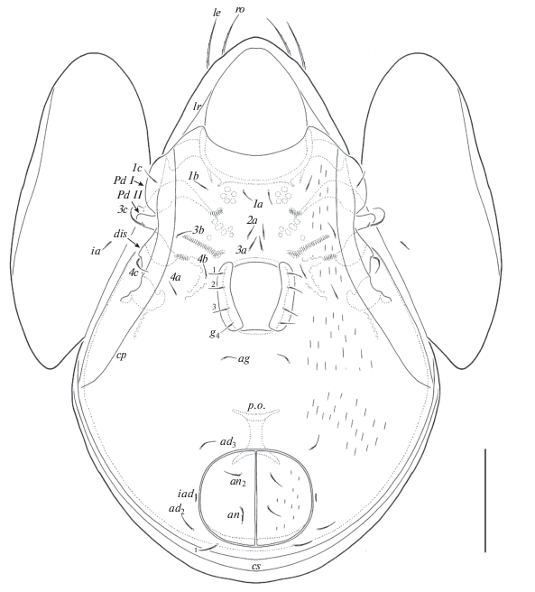
Fig. 3.
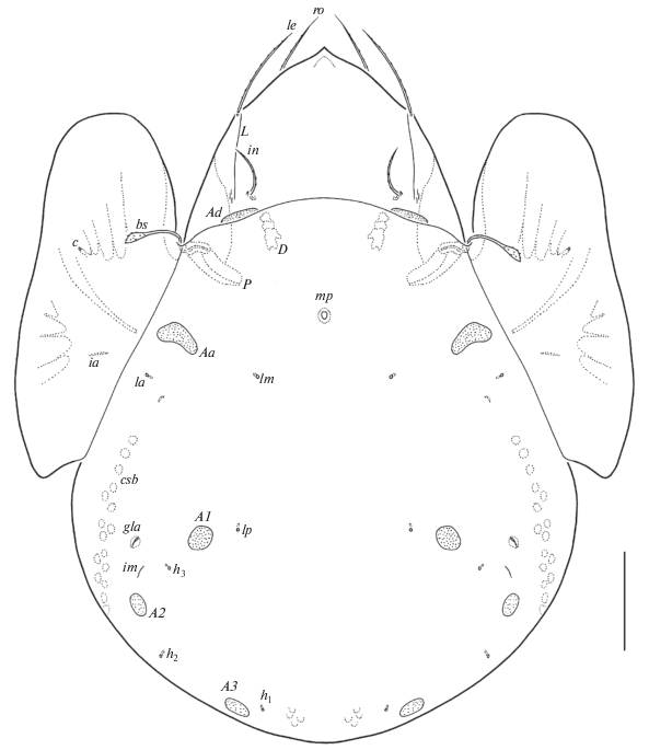
Neoribates (Neoribates) yangensis Ermilov sp. n., adult: 1 – anterior part of body, lateral view (pteromorph, gnathosoma and legs not shown); 2 – posterior part of body, lateral view; 3 – posterior view. Scale bar: 100 μm.
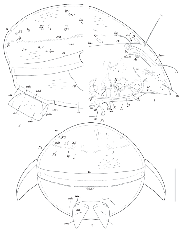
Fig. 4.
Neoribates (Neoribates) yangensis Ermilov sp. n., adult: Neoribates (Neoribates) yangensis Ermilov sp. n., adult: 1 – subcapitulum, ventral view; 2 – palp, right, antiaxial view; 3 – chelicera, right, antiaxial view. Scale bar (µm): 1, 3 – 17; 2 – 15.
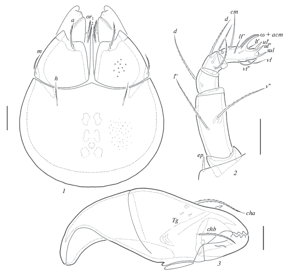
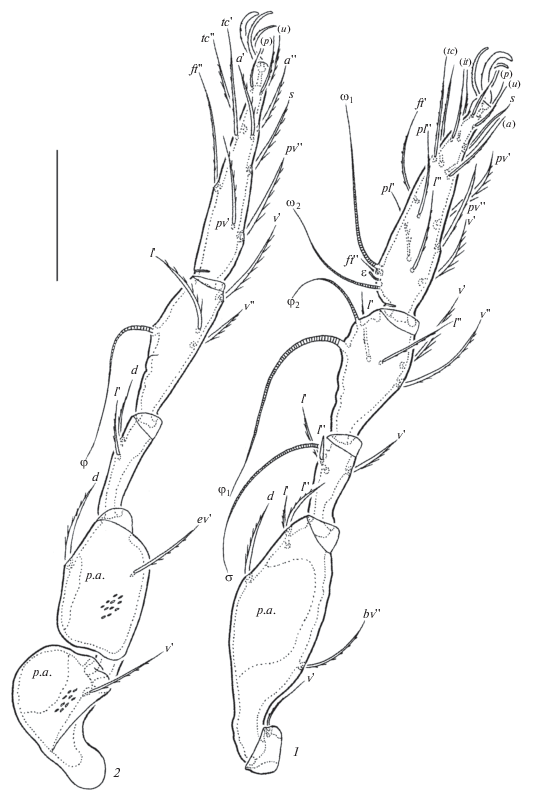
Fig. 5.
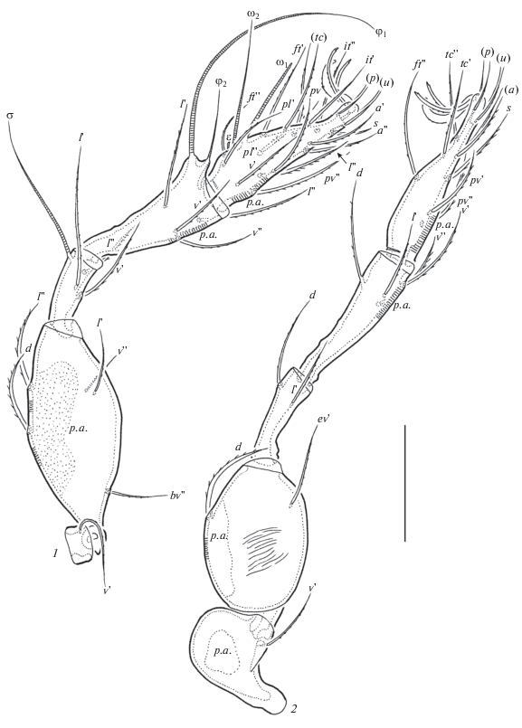
Neoribates (Neoribates) yangensis Ermilov sp. n., adult: 1 – leg I, left, paraxial view; 2 – leg IV, left, antiaxial view. Scale bar: 50 μm.
Material. The holotype (♀) and 6 paratypes (2 ♀♀, 4 ♂♂): Taiwan, Taipei City, Beitou Dist., Shamao Rd., Yangmingshan National Park, 25°8.573′ N, 121°32.607′ E, 352 m a.s.l., sample #16, lichen and soil, 17.IX.2017, collected by J.-R. Liao and H.C. Lee.
The holotype (ethanol with drop of glycerol) is deposited in NTU; six paratypes (ethanol with drop of glycerol) are deposited in TSUMZ.
Diagnosis. Body size: 464–564 × 298–381. Notogaster, anogenital region and lateral parts of epimeres lineolate. Rostrum rounded. Rostral, lamellar and interlamellar setae long, setiform, barbed. Bothridial setae, lanceolate, barbed. Notogastral setae short, thin, smooth. Saccules with small openings and channels. Subcapitular setae setiform, barbed, a longest and thickest, h shortest and thinnest. Epimeral and anogenital setae short, thin, slightly barbed. Four pairs of genital setae. Adanal setae ad3 inserted anterolateral to anal aperture. Leg tarsi with three claws.
Description. Measurements. Body length: 564 (holotype), 464–564 (6 paratypes); notogaster width: 381 (holotype), 298–381 (6 paratypes). Females larger than males: 547–564 × 365–381 versus 464–531 × 298–348.
Integument (Figs 1; 2; 3, 1–3; 4, 1). Body color brown. Body surface microfoveolate (visible under high magnification, ×1000). Rostrum sparsely foveolate (diameter of foveoles up to 4). Surface of notogaster, anogenital region, anal plates and lateral parts of epimeres lineolate, with short (length up to 16) depressed, distinct lines. All leg femora and trochanters III, IV with elongate tubercles or thin ridges antiaxially.
Prodorsum (Figs 1; 3, 1). Rostrum rounded. Lamellae located dorsolaterally, about half of prodorsum length (measured in lateral view), their ends slightly curving anteromedially. Translamellar line absent. Sublamellae slightly shorter than lamellae. Sublamellar porose areas rounded (12–14), partially covered by sublamellae. Prodorsal setae strongly varies in length in different specimens; rostral (53–57 up to 86), lamellar (61–65 up to 114) and interlamellar (94–98 up to 123) setae setiform, barbed, le inserted on the lamellae ends, bothridial setae (53–57 up to 90) with long stalk and shorter, lanceolate, barbed head. Exobothridial setae and their alveoli absent. Sejugal porose areas (36–41 × 4–6) narrowly elongated. Anterior, posterior and lateral ridges simple.
Notogaster (Figs 1; 3, 1–3). Anterior notogastral margin convex medially. Ten pairs of notogastral setae short (4–6), thin, smooth. Four pairs of saccules with small openings and channels. Setae lp inserted medial and close to S1. All lyrifissures and opisthonotal gland openings well visible, im located posterior to lm, ip between p1 and p2, ih and ips in lateral position, distanced from each other, gla posterior to im. Circumgastric scissure and circumgastric sigillar band distinct.
Gnathosoma (Fig. 4, 1–3). Subcapitulum longer than wide (123–135 × 90–98). Subcapitular setae setiform, barbed, a (20–24) longer and thicker than m (16–20) and h (12–16). Adoral setae (16–20) setiform, heavily barbed. Palps (69–73) with typical setation 0–2–1–3–9(+1 solenidion). Postpalpal setae (6) spiniform, smooth. Chelicerae (123–135) with two setifrom, barbed setae, cha (41–49) longer than chb (28–32). Trägårdh’s organ of chelicerae elongate triangular.
Epimeral and lateral podosomal regions (Figs 2; 3, 1, 2). Epimeral setal formula 3–1–3–3. Setae setiform, thin, slightly barbed, 3с (32) longer than others (20); 2a and 3a thickest. Pedotecta II rectangular, rounded in ventral view. Discidia triangular. Circumpedal carinae long, reaching anterior margin of ventral plate.
Anogenital region (Figs 2; 3, 1–3). Four pairs of genital, one pair of aggenital, two pairs of anal and three pairs of adanal setae similar in length (20), setiform, slightly barbed. Setae ad3 inserted anterolateral and close to anal aperture. Adanal lyrifissures located close and parallel to anal plates. Marginoventral porose area represented by numerous rounded and elongate oval parts.
Legs (Fig. 5, 1, 2). Tridactylous, median claw thicker than laterals, all serrated on dorsal side. Lateral claws with one slightly developed tooth ventrodistally. Dorsoparaxial porose areas on femora and trochanters III, IV, posteroventral porose areas on tarsi and anteroventral porose areas on tibiae well visible. Formulas of leg setation and solenidia: I (1–5–3–4–20) [1–2–2], II (1–5–3–4–15) [1–1–2], III (2–3–1–3–15) [1–1–0], IV (1–2–2–4–12) [0–0–0]; homologies of setae and solenidia indicated in Table 1. Famulus of tarsi I short, erect, slightly dilated distally, inserted between solenidion ω2 and seta ft ''.
Table 1.
Leg setation and solenidia of Neoribates (Neoribates) yangensis Ermilov sp. n.
| Leg | Tr | Fe | Ge | Ti | Ta |
|---|---|---|---|---|---|
| I | v' | d, (l), bv'', v'' | (l), v', σ | (l), (v), φ1, φ2 | (ft), (tc), (it), (p), (u), (a), s, (pv), v', (pl), l '', ɛ, ω1, ω2 |
| II | v' | d, (l), bv'', v'' | (l), v', σ | (l), (v), φ | (ft), (tc), (it), (p), (u), (a), s, (pv), ω1, ω2 |
| III | l ', v' | d, l ', ev' | l ', σ | l ', (v), φ | (ft), (tc), (it), (p), (u), (a), s, (pv) |
| IV | v' | d, ev' | d, l ' | d, l ', (v) | ft '', (tc), (p), (u), (a), s, (pv) |
Differential diagnosis. Neoribates (Neoribates) yangensis sp. n. is morphologically most similar to N. (N.) similis Fujikawa 2007 from Japan (Fujikawa, 2017) and N. (N.) pyramidalis (Tseng 1984) from Taiwan (Tseng, 1984) in having rounded rostrum, lanceolate bothridial setae, minute notogastral setae, four pairs of genital setae and localization of adanal setae ad3 in preanal position, but differs by the lineolate body surface (versus lineolate ornamentation absent).
Etymology. The specific name yangensis refers to the [Yang] mingshan National Park, where the new species was found.
Galumna (Galumna) tsengi Ermilov et Liao 2017 (Fig. 6)
Fig. 6.
Galumna (Galumna) tsengi Ermilov et Liao 2017, adult: dorsal view (legs not shown). Scale bar: 100 μm.
Material. Eight specimens (5 ♀♀, 3 ♂♂): Taiwan, Nantou County, Lugu Township, Fenghuang Nature Education Area, 23°43.735′ N, 120°47.287′ E, 875 m a.s.l., sample #5, soil and debris from tea tree, 6.VII.2017, collected by J.-R. Liao.
All specimens (ethanol with drop of glycerol) are deposited in TSUMZ.
Remarks. Galumna tsengi was described from soil of the Fushan Botanical Garden of Taiwan by Ermilov and Liao (2017a). Some specimens (2 females, 1 male) of this species from new materials (see Material section) from Taiwan are identical to the original description, but the other specimens (3 females, 2 males) with important difference: median pore located anterior to the virtual line between notogastral porose areas Aa (Fig. 6) versus posterior to the virtual line between A1 (Ermilov, Liao, 2017a). Hence, the variable localization of median pore in G. tsengi should be considered in future identification of this species.
Galumna (Galumna) alata (Hermann 1804) (Figs 7–11)
Material. Nine specimens (5 ♀♀, 4 ♂♂): Taiwan, New Taipei City, Tamsui Dist., Balaka Highway, Yangmingshan National Park, 25°10.969′ N, 121°30.564′ E, 703 m a.s.l., sample #20, soil, 17.IX.2017, collected by J.-R. Liao and H.C. Lee.
All specimens (ethanol with drop of glycerol) are deposited in TSUMZ.
Supplementary description (based on specimens from Taiwan). Measurements. Body length: 381–431 (9 specimens); notogaster width: 249–298 (9 specimens). Females larger than males: 415–431 × 282–298 versus 381–398 × 249–265.
Integument. Body color brown. Body surface microfoveolate (visible under high magnification, ×1000). All leg femora and trochanters III, IV with elongate tubercles antiaxially.
Fig. 7.
Galumna (Galumna) alata (Hermann 1804), adult: dorsal view (legs not shown). Scale bar: 100 μm.
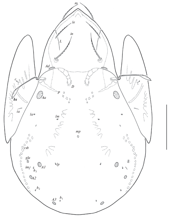
Fig. 8.
Galumna (Galumna) alata (Hermann 1804), adult: ventral view (gnathosoma and legs not shown). Scale bar: 100 μm.
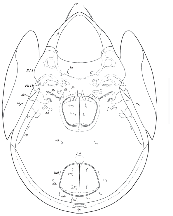
Fig. 9.
Galumna (Galumna) alata (Hermann 1804), adult: 1 – anterior part of body, lateral view (pteromorph, gnathosoma and legs not shown); 2 – posterior part of body, lateral view; 3 – posterior view. Scale bar: 100 μm.
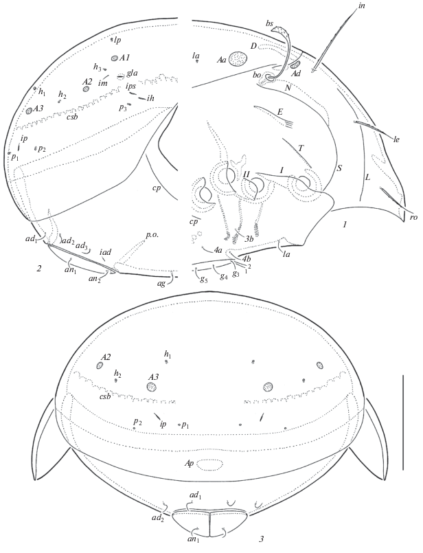
Fig. 10.
Galumna (Galumna) alata (Hermann 1804), adult: 1 – subcapitulum, ventral view; 2 – palp, right, antiaxial view; 3 – chelicera, left, paraxial view. Scale bar (µm): 1, 3 – 17; 2 – 15.
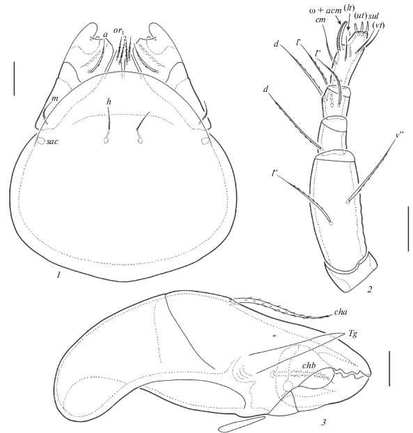
Fig. 11.
Galumna (Galumna) alata (Hermann 1804), adult: 1 – leg I, right, antiaxial view; 2 – leg IV, left, antiaxial view. Scale bar: 50 μm.
Prodorsum (Figs 7; 9, 1). Rostrum pointed. Lamellar and sublamellar lines thin, slightly divergent distally, S curving backwards at ventral ends, L directed to lateral sides of prodorsum. Prodorsal leg niches and lateral ridges of prodorsum well-developed. Rostral (45–53), lamellar (41–49) and interlamellar (69–77) setae setifrom, barbed. Bothridial setae (77–86) with long, smooth stalks and short, lanceolate, barbed heads. Exobothridial setae and their alveoli absent. Sejugal porose areas (12–14 × 4–6) elongate oval, transversely orientated, posterolateral to in. Dorsophragmata elongated, represented by group of low, roughened thickenings collectively elongated longitudinally, with variable number of components.
Notogaster (Figs 7; 9, 1–3). Dorsosejugal suture complete, but very poorly visible medially. With 10 pairs of setal alveoli and four pairs of rounded porose areas, Aa (18–20) larger than others (8–10). Setal alveoli la located close to the pteromorphal hinges, posterior to Aa. Median pore present, represented by one part, located clearly anterior to the virtual line between A1. All lyrifissures distinct, im located posterolateral to Aa, ip between p1 and p1, ih and ips close to each other, anterior to p3. Opisthonotal gland openings located lateral to A1.
Gnathosoma (Fig. 10, 1–3). Subcapitulum longer than wide (123–135 × 98–106). Subcapitular setae setiform, slightly barbed, a (20–24) longer and thicker than m (16–20) and h (14–18). Adoral setae (16–20) setiform, heavily barbed. Palps (90–94) with typical setation 0–2–1–3–9(+1 solenidion). Postpalpal setae (4) spiniform, smooth. Chelicerae (155–164) with two setifrom, barbed setae, cha (49–57) longer than chb (28–36). Trägårdh’s organ of chelicerae elongate triangular.
Epimeral and lateral podosomal regions (Figs 8; 9, 1, 2). Anterior tectum of epimere I smooth. Pedotecta I and II rounded in ventral view. Discidia triangular. Epimeral setal formula: 1–0–1–2. Epimeral setae (10–14) setiform, thin, slightly barbed. Circumpedal carinae thin, directed to insertions of 3b, but clearly not reaching them.
Anogenital region (Figs 8; 9, 1–3). Six pairs of genital (g1, g2, 12–16; g3–g6, 8–10), one pair of aggenital (8–10), two pairs of anal (8–10) and three pairs of adanal (8–10) setae setiform, thin, slightly barbed. Anterior edges of genital plates with three setae. Aggenital setae located between genital and anal apertures, equal distanced from them. Adanal lyrifissures located close and parallel to anal plates. Adanal setae ad1 and ad2 postanal, ad3 paraanal and posterolateral to iad. Distance ad1–ad2 = ad2–ad3. Unpaired postanal porose area elongate oval (24–26 × 6–10).
Legs (Fig 11, 1, 2). Median claw distinctly thicker than laterals, all barbed on dorsal sides. Dorsoparaxial porose areas on all femora and on trochanters III, IV well visible. Formulas of leg setation and solenidia: I (1–4–3–4–20) [1–2–2], II (1–4–3–4–15) [1–1–2], III (1–2–1–3–15) [1–1–0], IV (1–2–2–3–12) [0–1–0]; homologies of setae and solenidia indicated in Table 2. Solenidion of tibiae IV inserted in medioanterior part of the segments. Famulus of tarsi I short, erect, slightly dilated distally, inserted posterior to solenidia ω1.
Table 2.
Leg setation and solenidia of Galumna (Galumna) alata (Hermann 1804)
| Leg | Tr | Fe | Ge | Ti | Ta |
|---|---|---|---|---|---|
| I | v' | d, (l), bv'' | (l), v', σ | (l), (v), φ1, φ2 | (ft), (tc), (it), (p), (u), (a), s, (pv), v', (pl), l'', ɛ, ω1, ω2 |
| II | v' | d, (l), bv'' | (l), v', σ | (l), (v), φ | (ft), (tc), (it), (p), (u), (a), s, (pv), ω1, ω2 |
| III | v' | d, ev' | l ', σ | l ', (v), φ | (ft), (tc), (it), (p), (u), (a), s, (pv) |
| IV | v' | d, ev' | d, l ' | l ', (v), φ | ft'', (tc), (p), (u), (a), s, (pv) |
See Table 1 for explanations.
Remarks. Galumna alata has a semicosmopolitan distribution (Subías, 2004, updated 2018). An adult of this species was described by Hermann (1804); it was later redescribed and illustrated by several authors: e.g., Oudemans (1917), Jacot (1934), Grandjean (1936), Bayartogtokh (2010), Seniczak et al. (2012). The specimens of G. alata from Taiwan are similar in general appearance to those from redescriptions according to the following traits: rostrum pointed; lamellar and sublamellar lines slightly divergent distally; bothridial setae lanceolate; rostral, lamellar and interlamellar setae long, in longest; notogastral suture present; four pair of rounded notogastral porose areas; setal alveoli la inserted posterior to Aa; im located lateral or posterolateral to A1; anogenital setae short; postanal porose area oval; surface without sculpturing and ornamentation. However, some differences in the morphology and localization of median pore are present: a) median pore is represented by one part (versus several parts or absent in redescriptions); b) median pore located clearly anterior to the virtual line, between notogastral porose areas A1 (Fig. 7), (versus being posterior (if present) to the virtual line between A1 in redescriptions). Hence, the variable morphology and localization of median pore in G. alata should be considered in the future identification of this species.
Список литературы
Aoki J., 1991. Oribatid mites of high altitude forests of Taiwan I. Mt. Pei-ta-wu Shan // Acta Arachnologica. V. 40. P. 75–84.
Bayartogtokh B., 2010. Oribatid mites of Mongolia (Acari: Oribatida). М.: КМК. 372 p.
Chu Y.I., Aoki J., 1997. Fauna of oribatid mite at Fu-shan forest litter and humus layer // Chinese Journal of Entomology. V. 17. № 3. P. 172–178.
Ermilov S.G., 2018. Contribution to the knowledge of the oribatid mite genus Aokiella (Acari, Oribatida, Carabodidae) // Systematic and Applied Acarology. V. 23. № 4. P. 643–651.
Ermilov S.G., Corpuz-Raros L., 2015. A new subgenus and two new species of oribatid mites of the genus Neoribates (Acari, Oribatida, Parakalummidae) from the Philippines // Zootaxa. V. 3956. № 2. P. 224–238.
Ermilov S.G., Klimov P.B., 2017. Generic revision of the large-winged mite superfamily Galumnoidea (Acari, Oribatida) of the world // Zootaxa. V. 4357. № 1. P. 1–72.
Ermilov S.G., Liao J.-R., 2017. Neotype designations and supplementary descriptions of Taiwanoppia subtropica Tseng, 1982 and Muliercula chiayiensis Tseng, 1984 (Acari: Oribatida: Oppiidae, Scheloribatidae) // Systematic and Applied Acarology. V. 22. № 7. P. 897–914.
Ermilov S.G., Liao J.-R., 2017a. New faunistic and taxonomic data on oribatid mites (Acari, Oribatida) of Taiwan // Systematic and Applied Acarology. V. 22. № 6. P. 824–840.
Ermilov S.G., Starý J., 2018. Oribatid mites (Acari, Oribatida) from Tam Dao National Park (Northern Vietnam), with description of two new species // Systematic and Applied Acarology. V. 23. № 2. P. 314–338.
Grandjean F., 1936. Les Oribates de Jean Frédéric Hermann et de son père // Annales de la Société Entomologique de France. V. 105. P. 27–110.
Fujikawa T., 2007. Two new species of Neoribates (Neoribates) (Acari, Oribatida) from Shikoku Island, Japan // Edaphologia. V. 81. P. 1–7.
Jacot A.P., 1934. The Galumnas (Oribatoidea – Acarina) of the Northeastern United States // Journal of the New York Entomological Society. V. 42. № 1. P. 87–118.
Hermann J.F., 1804. Memoire apterologique. Strassbourg. P. 1–144.
Oudemans A.C., 1917. Notizen über Acari, 26. Reihe (Oribatoidea) (Gruppe der Galumnae) // Archiv für Naturgeschichte (A). V. 83. № 4. P. 1–84.
Seniczak S., Iturrondobeitia J.C., Seniczak A., 2012. The ontogeny of morphological traits in three species of Galumnidae (Acari: Oribatida) // International Journal of Acarology. V. 38. № 7. P. 612–638.
Subías L.S., 2004. Listado sistemático, sinonímico y biogeográfico de los ácaros oribátidos (Acariformes: Oribatida) del mundo (excepto fósiles) // Graellsia. V. 60 (número extraordinario). P. 3–305. Online version accessed in January 2018. 605 p. http://bba.bioucm.es/cont/docs/RO_1.pdf
Tseng Y., 1984. Taxonomical study of oribatid mites from Taiwan (Acarina: Astigmata) (II) // Chinese Journal of Entomology. V. 4. P. 27–74.
Дополнительные материалы отсутствуют.
Инструменты
Зоологический журнал