Зоологический журнал, 2019, T. 98, № 9, стр. 1037-1047
Two New Species of Oripodoidea (Acari, Oribatida) from Ethiopia
S. G. Ermilov *
Tyumen State University
625003 Tyumen, Russia
* E-mail: ermilovacari@yandex.ru
Поступила в редакцию 10.10.2018
После доработки 13.01.2019
Принята к публикации 27.02.2019
Аннотация
Two new species of oribatid mites (Acari, Oribatida) of the superfamily Oripodoidea are described from Ethiopia. Pilobatella dhatiensis sp. n. differs from P. schauenbergi Mahunka, 1977 by the larger body and long, barbed lamellar and interlamellar setae. Muliercula walalensis sp. n. differs from all other species of the genus by the presence of a translamella.
During my taxonomic studies of oribatid mites (Acari, Oribatida) in the Ethiopian Dhati-Walal National Park, I discovered two new species of the superfamily Oripodoidea, one belonging to the genus Pilobatella Balogh et Mahunka 1967 (family Haplozetidae), and the other to Muliercula Coetzer 1968 (family Scheloribatidae). The primary goal of the paper is to describe and illustrate these new species.
Pilobatella was proposed by Balogh and Mahunka (1967) with Pilobatella punctulata Balogh et Mahunka 1967 as type species. Currently, the genus includes 10 species (Ermilov et al., 2012), which are distributed in the Ethiopian and Oriental regions, as well as in northern India. The main diagnostic characteristics were summarized by Balogh and Mahunka (1967) and Balogh and Balogh (1984, 1992), and augmented by Ermilov et al. (2012). An identification key to the known species of Pilobatella was presented by Ermilov et al. (2012).
Muliercula was proposed by Coetzer (1968) with Muliercula muliercula Coetzer 1968 as type species. Currently, the genus includes 10 species (Subías, 2004, updated 2018), which are registered in the Ethiopian, Neotropical and Oriental regions. The main diagnostic characteristics were summarized by Coetzer (1968). An identification key to the known species (except one) of Muliercula was presented by Shtanchaeva et al. (2014).
This work is part of my ongoing study of the oribatid mite fauna of Ethiopia (e.g., Ermilov et al., 2014; Ermilov, 2016; Ermilov and Rybalov, 2018).
METHODS
Litter and fallen leaves were collected by using a stainless steel frame (50 × 50 cm) with a sieve (mesh size 2 × 2 cm). Oribatid mites were extracted into 75% ethanol using Berlese funnels with electric bulbs (150 W) in laboratory conditions.
Specimens were mounted in lactic acid on temporary cavity slides for measurement and illustration. Body length was measured in lateral view, from the tip of the rostrum to the posterior edge of the notogaster. Notogastral width refers to the maximum width of the notogaster in dorsal view. Lengths of body setae were measured in lateral aspect. All body measurements are presented in micrometers. Formulas for leg setation are given in parentheses according to the sequence trochanter–femur–genu–tibia–tarsus (famulus included). Formulas for leg solenidia are given in square brackets according to the sequence genu–tibia–tarsus.
Drawings were made with a camera lucida using a Leica transmission light microscope “Leica DM 2500”.
Morphological terminology used in this paper follows that of F. Grandjean: see Travé and Vachon (1975) for references, Norton (1977) for leg setal nomenclature, and Norton and Behan-Pelletier (2009), for an overview.
The following abbreviations are used: lam – lamella; tlam – translamella; slam – sublamella; plam – prolamella; Al – sublamellar porose area; Sl – sublamellar saccule; tu – tutorium; lr – lateral ridge; vlr – ventrolateral ridge; ro, le, in, bs, ex – rostral, lamellar, interlamellar, bothridial and exobothridial setae, respectively; Ad – sejugal porose area; D – dorsophragma; P – pleurophragma; c, la, lm, lp, h, p – notogastral setae; Sa, S1, S2, S3, S4 – notogastral saccules; ia, im, ip, ih, ips – notogastral lyrifissures; gla – opisthonotal gland opening; h, m, a – subcapitular setae; or – adoral seta; v, l, d, cm, acm, ul, sul, vt, lt – palp setae; ω – palp and leg solenidion; Pd I, Pd II – pedotecta I, II, respectively; 1a, 1b, 1c, 2a, 3a, 3b, 3c, 4a, 4b, 4c – epimeral setae; dis – discidium; cp – circumpedal carina; g, ag, an, ad – genital, aggenital, anal and adanal setae, respectively; iad – adanal lyrifissure; p.o. – preanal organ; Tr, Fe, Ge, Ti, Ta – leg trochanter, femur, genu, tibia, tarsus, respectively; t – tooth; pr – process; ap – apophysis; p.a. – leg porose area; σ, φ – leg solenidia; ɛ – leg famulus; v, ev, bv, l, d, ft, tc, it, p, u, a, s, pv, pl – leg setae.
The following abbreviations of the relevant institutional collections are used: SMNH – Senckenberg Museum of Natural History, Görlitz, Germany; TSUMZ – Tyumen State University Museum of Zoology, Tyumen, Russia.
SYSTEMATICS
Genus Pilobatella Balogh et Mahunka 1967
Type species Pilobatella punctulata Balogh et Mahunka 1967.
Pilobatella dhatiensis Ermilov sp. n. (Figs 1–3)
Fig. 1.
Pilobatella dhatiensis sp. n., adult: a – dorsal view; b – anterior part of body (gnathosoma and legs not illustrated), lateral view; c – posterior part of body, lateral view; d – posterior view. Scale bar 100 µm.
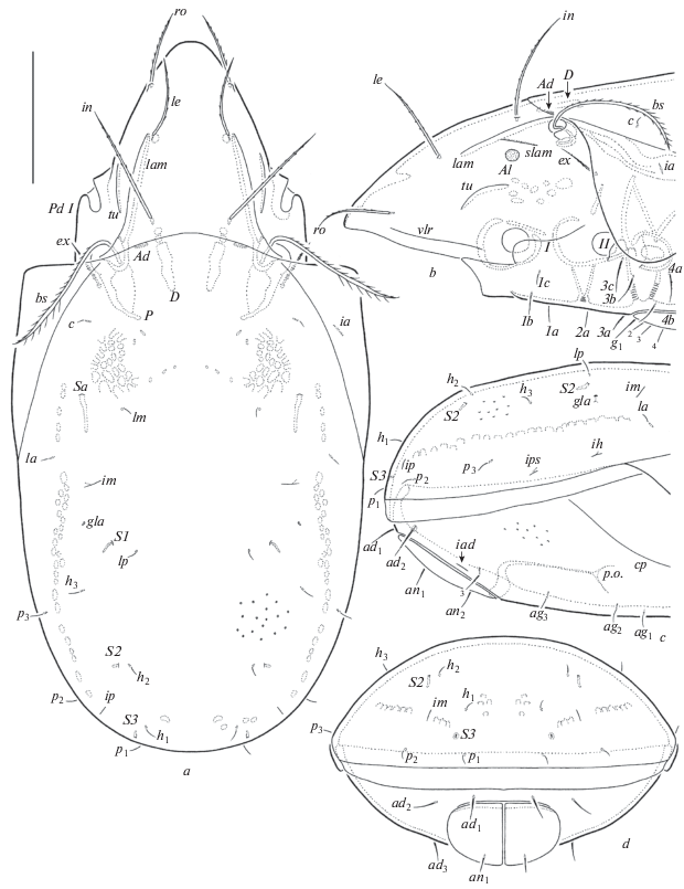
Fig. 2.
Pilobatella dhatiensis sp. n., adult: a – ventral view (gnathosoma and legs not illustrated); b – chelicera, left, paraxial view; c – subcapitulum, ventral view; d – palp, left, paraxial view. Scale bar (µm): a – 100; b–d – 20.
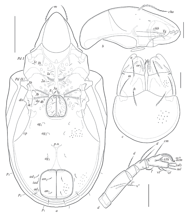
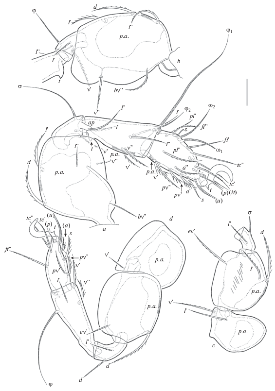
Fig. 3.
Pilobatella dhatiensis sp. n., adult: a – leg I, without trochanter and basal part of femur, right, antiaxial view; b – trochanter (covered by pedotectum II partially), femur, genu and basal part of tibia of leg II, left, antiaxial view; c – trochanter, femur and genu of leg III, right, antiaxial view; d – leg IV, left, antiaxial view. Scale bar: 20 µm.
Material. Holotype (♀) and five paratypes (3♀♀, 2♂♂): Ethiopia, Dhati-Walal National Park, 9°26′35.5″ N, 34°48′07.6″ E, rarefied forest with needle leaves bush, litter, 27.X.2017 (L.B. Rybalov). Three paratypes (1♀, 2♂♂): Ethiopia, Dhati-Walal National Park, 9°26′33.7″ N, 34°47′22.5″ E, mixed deciduous forest in the valley, litter, 27.X.2017 (L.B. Rybalov). Four paratypes (2♀♀, 2♂♂): Ethiopia, near Dhati-Walal National Park, 9°25′01.2″ N, 34°45′21.3″ E, mixed forest on the mountain, litter, 20.X.2017 (L.B. Rybalov).
The holotype (ethanol with a drop of glycerol) is deposited in SMNH; 12 paratypes (ethanol with a drop of glycerol) are deposited in TSUMZ.
Diagnosis. Body size: 531–614 × 249–298. Body surface microgranulate, notogaster and ventral side foveolate. Rostral, lamellar and interlamellar setae long, setiform, barbed; ro shortest, in longest. Bothridial setae thickened, ciliate. Exobothridial setae of medium size, setiform, erect, barbed. Notogastral setae short, setiform, smooth. Saccules Sa with a very long channel, other saccules with a slightly elongate channel. Epimeral and anogenital setae setiform, slightly barbed. Leg tarsi I with 19 setae (l '' absent). Leg setae l '' on genua I inserted on an elongate apophysis.
Description. Measurements. Body length: 531 (holotype), 531–614 (paratypes); notogaster width: 257 (holotype), 249–298 (paratypes). No difference between females and males in body size.
Integument. Body color brown. Body surface densely microgranulate (visible under high magnification). Notogaster, ventral side, anal plates and subcapitular mentum and genae sparsely foveolate (diameter of foveoles up to 4).
Prodorsum (Figs 1a, 1b). Rostrum broadly rounded. Lamellae located dorsolaterally, half as long as prodorsum. Translamellar and prolamellar lines absent. Sublamellae thin, half as long as lamellae. Sublamellar porose areas rounded (10–16) or oval (10–16 × 8–12). Tutoria and ventrolateral ridges well-developed. Rostral (49–57), lamellar (65–73) and interlamellar (73–86) setae setiform, barbed; le inserted medial and close to lamellar ends. Bothridial setae (102–114) thickened, ciliate. Dorsosejugal porose areas elongate oval (6–8 × 2–4). Exobothridial setae (36–41) setiform, erect, barbed.
Notogaster (Figs 1, 2a). Anterior notogastral margin slightly convex medially. Ten pairs of notogastral setae short (10–12), setiform, thin, smooth. Four pairs of saccules developed, Sa with a very long channel, other saccules (S1, S2, S3) with a slightly elongate channel. Distance S1–S1 slightly longer than S2–S2. Setae lm inserted medial to Sa, lp medial to S1. All lyrifissures distinct, im located between Sa and S1, ip posterolateral to h1, ih and ips in lateral position, anterior to p3. Opisthonotal gland openings located posterior to im. Circumgastric scissure and circumgastric sigillar band distinct.
Gnathosoma (Figs 2b–2d). Subcapitulum longer than wide (110–114 × 82–86). Subcapitular setae (a, m, h) similar in length (22–24), setiform, slightly barbed; m slightly thinner than others. Two pairs of adoral setae (12) setiform, heavily barbed. Palps (length 73–77) with setation 0–2–1–3–9(+ω). Postpalpal setae (6) spiniform, hardly barbed. Chelicerae (length 127–131) with two setiform, barbed setae, cha (41–45) longer than chb (26–28). Trägårdh’s organ of chelicerae elongate triangular, rounded distally.
Epimeral and lateral podosomal regions (Figs 1b, 2a). Epimeral setal formula: 3–1–3–3. Epimeral setae 3с and 4с (20–24) longer than 1b (16) and others (10–12), all setiform, slightly barbed; 3a slightly thicker than others. Pedotecta II trapezoid in ventral view. Discidia elongate triangular, rounded distally. Circumpedal carinae comparatively short, reaching level of insertions of acetabula IV.
Anogenital region (Figs 1b–1d, 2a). Six pairs of genital (g1, 20; g2–g6, 10–12), three pairs of aggenital (10–12), two pairs of anal (16) and three pairs of adanal (ad1, ad2, 20–24; ad3, 16) setae setiform, slightly barbed. Adanal lyrifissures located close and parallel to anal plates. Adanal setae ad1 in posterior, ad2 in posterolateral, ad3 in anterolateral positions; ad3 anterior to iad. Marginal porose area absent. Preanal organ goblet-like. Ovipositor elongated (151 × 41), blades (61) shorter than length of distal section (beyond middle fold; 90). Each of the three blades with four smooth setae, ψ1 ≈ τ1 (28) setiform, ψ2 ≈ τa ≈ τb ≈ τc (12) thorn-like. Six coronal setae spiniform (4).
Legs (Fig. 3). Claw of all tarsi strong, serrate on dorsal side, with tooth ventrobasally. Tibiae of leg I and II with triangular tooth ventrobasally. Femora II slightly bilobed ventrally. Trochanters IV with triangular process dorsoanteriorly. Dorsoparaxial porose areas on femora I–IV and on trochanters III, IV well visible. Ventral porose areas in basal parts of tarsi and in distal parts of tibiae distinct only on leg I and II. Formulas of leg setation and solenidia: I (1–5–3–4–19) [1–2–2], II (1–5–3–4–15) [1–1–2], III (2–3–1–3–15) [1–1–0], IV (1–2–2–3–12) [0–1–0]; homology of setae and solenidia indicated in Table 1. Famulus inserted posterior to solenidion ω2. Setae l '' on genua I inserted on elongate apophysis.
Table 1.
Leg setation and solenidia of adult Pilobatella dhatiensis sp. n. and Muliercula walalensis sp. n.
| Leg | Tr | Fe | Ge | Ti | Ta |
|---|---|---|---|---|---|
| I | v' | d, (l), bv'', v'' | (l), v', σ | (l), (v), φ1, φ2 | (ft), (tc), (it), (p), (u), (a), s, (pv), (pl), v', ɛ, ω1, ω2 |
| II | v' | d, (l), bv'', v'' | (l), v'*, σ | (l), (v), φ | (ft), (tc), (it), (p), (u), (a), s, (pv), ω1, ω2 |
| III | l ', v' | d, l ', ev' | l ', σ | l ', (v), φ | (ft), (tc), (it), (p), (u), (a), s, (pv) |
| IV | v' | d, ev' | d, l ' | l ', (v), φ | ft '', (tc), (p), (u), (a), s, (pv) |
Roman letters refer to normal setae, Greek letters – to solenidia (except ɛ = famulus). Single prime (') marks setae on the anterior and double prime ('') – setae on the posterior side of a given leg segment. Parentheses refer to a pair of setae. * – Setae v' on genua II absent in M. walalensis sp. n.
Remarks. Pilobatella dhatiensis sp. n. is similar to P. schauenbergi Mahunka 1977 from the Oriental region (see Mahunka, 1977) in the presence of saccules Sa with long channels. However, the former species differs from the latter by larger body size (531–614 × × 249–298 versus 348–407 × 188–212); also, lamellar and interlamellar setae are long and barbed in P. dhatiensis sp. n. (versus short and heavily ciliated in P. schauenbergi).
Etymology. The specific name dhatiensis refers to the Ethiopian Dhati-Walal National Park, where the type material was collected.
Muliercula walalensis Ermilov sp. n. (Figs 4–6)
Fig. 4.
Muliercula walalensis sp. n., adult: a – dorsal view; b – anterior part of body (gnathosoma and legs not illustrated), lateral view; c – posterior part of body, lateral view; d – posterior view. Scale bar 50 µm.
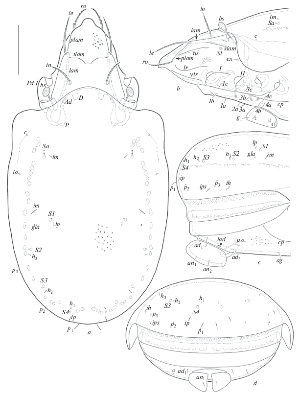
Fig. 5.
Muliercula walalensis sp. n., adult: a – ventral view (gnathosoma and legs not illustrated); b – chelicera, left, paraxial view; c – subcapitulum, ventral view; d – palp, right, antiaxial view. Scale bar (µm): a – 50; b–d – 20.
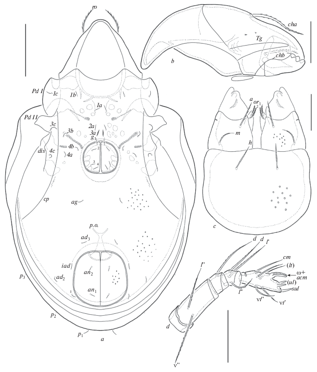
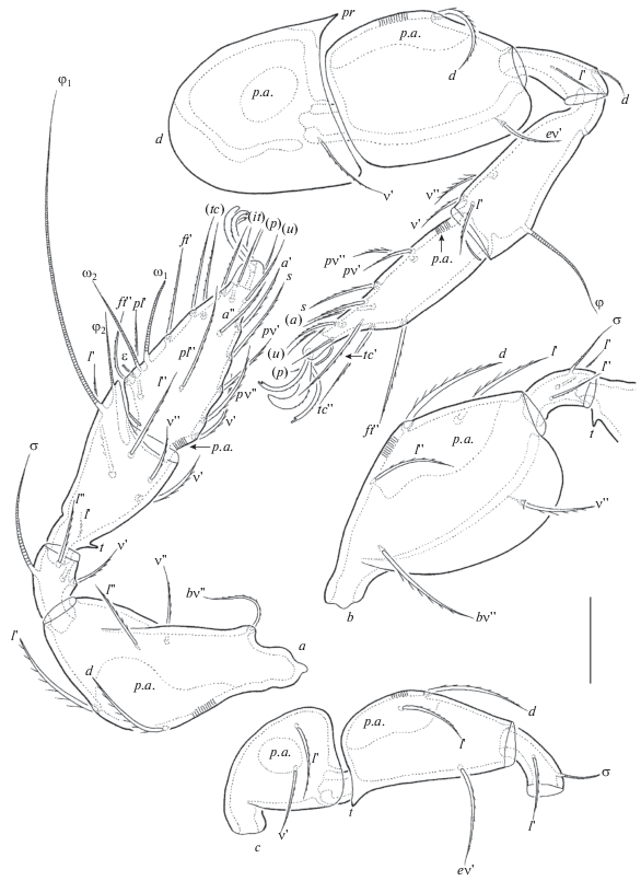
Fig. 6.
Muliercula walalensis sp. n., adult: a – leg I, without trochanter, right, antiaxial view; b – femur, genu and basal part of tibia of leg II, right, antiaxial view; c – trochanter, femur and genu of leg III, left, antiaxial view; d – leg IV, left, antiaxial view. Scale bar 20 µm.
Material. Holotype (♂) and six paratypes (4♀♀, 2♂♂): Ethiopia, Dhati-Walal National Park, 9°26′35.5″ N, 34°48′07.6″ E, rarefied forest with needle leaves bush, litter, 27.X.2017 (L.B. Rybalov). Twelve paratypes (9♀♀, 3♂♂): Ethiopia, Dhati-Walal National Park, 9°26′34.6″ N, 34°48′02.1″ E, bamboo forest, litter, 20.X.2017 (L.B. Rybalov).
The holotype (ethanol with drop of glycerol) is deposited in SMNH; 18 paratypes (ethanol with drop of glycerol) are deposited in TSUMZ.
Diagnosis. Body size: 298–381 × 166–215. Translamella developed, shortly interrupted medially. Prolamellae complete. Tutoria long. Rostral, lamellar and interlamellar setae long, setiform, barbed; le longest. Bothridial setae clavate, barbed. Notogastral setae p1 short, setiform, smooth, other setae vestigial. Five pairs of saccules present. Epimeral and anogenital setae setiform, slightly barbed. Leg tarsi I with 19 setae (l '' absent).
Description. Measurements. Body length: 332 (holotype), 298–381 (paratypes); notogaster width: 174 (holotype), 166–215 (paratypes). Females larger than males: 348–381 × 199–215 versus 298–332 × × 166–182.
Integument. Body color light brown to dark brown. Body surface densely microfoveolate (visible only in dissected specimens under high magnification). Notogaster, ventral side, anal plates, subcapitular mentum and genae and anterior part of prodorsum sparsely foveolate (diameter of foveoles up to 2). Podosomal regions microgranulate.
Prodorsum (Figs 4a, 4b). Rostrum slightly protruding, rounded. Lamellae located dorsolaterally, as long as half of prodorsum. Translamella long, shortly interrupted medially. Prolamellae present, complete. Sublamellae thin, half as long as lamellae. Sublamellar saccules present instead of porose areas. Tutoria and ventrolateral ridges well-developed. In addition, lateral ridges present between tutoria and ventrolateral ridges. Rostral (28–36), lamellar (45–53) and interlamellar (36–45) setae setiform, barbed. Bothridial setae (24–32) clavate, barbed. Dorsosejugal porose areas elongate oval (8 × 4). Exobothridial setae (8–12) setiform, slightly barbed.
Notogaster (Figs 4, 5a). Anterior notogastral margin slightly convex medially. Ten pairs of notogastral setae developed, p1 (8–12) setiform, thin, smooth, other setae (1) vestigial. Five pairs of saccules with drop-like channels. Distance S1–S1 shorter than S2–S2. Setae lm inserted medial to Sa, lp posteromedial to S1. All lyrifissures distinct, im located anterolateral to S1, ip lateral to h1, ih and ips in lateral position, ih anterolateral to p3, ips posterolateral to p3. Opisthonotal gland openings located posterior to im. Circumgastric scissure and circumgastric sigillar band distinct.
Gnathosoma (Figs 5b–5d). Subcapitulum longer than wide (69–77 × 49–57). Subcapitular setae a and h (12–14) longer than m (8–10), all setiform, barbed. Two pairs of adoral setae (6) setiform, heavily barbed. Palps (length 45–49) with setation 0–2–1–3–9(+ω). Postpalpal setae (4) spiniform, smooth. Chelicerae (length 69–77) with two setiform, barbed setae, cha (24–28) longer than chb (16–18). Trägårdh’s organ of chelicerae elongate triangular, rounded distally.
Epimeral and lateral podosomal regions (Figs 4b, 5a). Epimeral setal formula: 3–1–3–3. Epimeral setae 3b, 3с and 4с (12–16) longer than others (8–12), all setiform, slightly barbed; 3a slightly thicker than others. Pedotecta II trapezoid in ventral view. Discidia elongate triangular, rounded distally. Circumpedal carinae long, reaching level of pedotecta II.
Anogenital region (Figs 4b–4d, 5a). Four pairs of genital (g1, 8–12; g2–g4, 6–8), one pair of aggenital (8–12), two pairs of anal (8–12) and three pairs of adanal (8–12) setae setiform, slightly barbed. Adanal lyrifissures located close and parallel to anal plates. Adanal setae ad1 in posterolateral, ad2 in lateral, ad3 in anterior positions; ad3 anterior to iad. Marginal porose area absent. Preanal organ goblet-like. Ovipositor elongated (122 × 32), blades (57) shorter than length of distal section (beyond middle fold; 65). Each of the three blades with four smooth setae, ψ1 ≈ τ1 (24) setiform, ψ2 ≈ τa ≈ τb ≈ τc (10–12) thorn-like. Six coronal setae spiniform (2).
Legs (Fig. 6). Median claw distinctly thicker than laterals, all serrate on dorsal side; lateral claws with a very small tooth ventrodistally. Tibiae of leg I and II each with a triangular tooth ventrobasally. Femora III each with a triangular tooth ventrobasally. Dorsoparaxial porose areas on femora I–IV and on trochanters III, IV well visible. Ventral porose areas in basal parts of tarsi present, ventral porose areas in distal parts of tibiae absent. Trochanters IV each with a triangular process dorsoanteriorly. Formulas of leg setation and solenidia: I (1–5–3–4–19) [1–2–2], II (1–5–2–4–15) [1–1–2], III (2–3–1–3–15) [1–1–0], IV (1–2–2–3–12) [0–1–0]; homology of setae and solenidia indicated in Table 1. Famulus inserted posterior to solenidion ω2.
Remarks. Muliercula walalensis sp. n. differs from all other species of the genus by the presence of a well-developed translamella (versus translamella being absent in others).
Etymology. The specific name walalensis refers to the Ethiopian Dhati-Walal National Park, where the type material was collected.
Список литературы
Balogh J., Balogh P., 1984. A review of the Oribatuloidea Thor, 1929 (Acari: Oribatei) // Acta Zoologica Hungarica. V. 30. № 3–4. P. 257–313.
Balogh J., Balogh P., 1992. The oribatid mites genera of the World. V. 1. Budapest: Hungarian National Museum Press. 263 p.
Balogh J., Mahunka S., 1967. The scientific results of the Hungarian soil zoological expeditions to the Brazzaville-Congo. 30. The oribatid mites (Acari) of Brazzaville-Congo, II // Opuscula Zoologica Budapest. V. 7. № 1. P. 35–43.
Coetzer A., 1968. New Oribatulidae Thor, 1929 (Oribatei, Acari) from South Africa, new combinations and a key to the genera of the family // Memórias do Instituto de Investigaçāo Cientifica de Moçambique. V. 9 (A). P. 15–126.
Ermilov S.G., 2016. Additions to the oribatid mite fauna of Central Ethiopia, with description of a new species of Scheloribates (Bischeloribates) // Spixiana. V. 39. № 1. P. 75–82.
Ermilov S.G., Rybalov L.B., 2018. New faunistic and taxonomic data on oribatid mites (Acari, Oribatida) of Ethiopia // Systematic and Applied Acarology. V. 23. № 9. P. 1827–1837.
Ermilov S.G., Rybalov L.B, Hundama T., 2014. Ethiopian oribatid mites (Acari, Oribatida): results of the Joint Russian-Ethiopian Biological Expedition (June 2013) // Systematic and Applied Acarology. V. 19. № 2. P. 197–204.
Ermilov S.G., Winchester N.N., Lowman M.M., Wassie A., 2012. Two new species of oribatid mites (Acari: Oribatida) from Ethiopia, including a key to species of Pilobatella // Systematic and Applied Acarology. V. 17. № 3. P. 301–317.
Mahunka S., 1977. Neue und interessante Milben aus dem Genfer Museum XX. Contribution to the oribatid fauna of S.E. Asia (Acari, Oribatida) // Revue suisse de Zoologie. V. 84. № 1. P. 247–274.
Norton R.A., 1977. A review of F. Grandjean’s system of leg chaetotaxy in the Oribatei (Acari) and its application to the family Damaeidae // Dindal D.L., ed. Biology of oribatid mites. Syracuse, SUNY College of Environmental Science and Forestry. P. 33–61.
Norton R.A., Behan-Pelletier V.M., 2009. Oribatida // A Manual of Acarology (TX): Lubbock, Texas Tech University Press. P. 430–564.
Shtanchaeva U.Ya., Ermilov S.G., Tolstikov A.V., Subías L.S., 2014. Supplementary description of Indoribates (Haplozetes) minutus (Tseng, 1984) and Muliercula femoroserrata (Pérez-Iñigo et Baggio, 1980) (Acari, Oribatida, Oripodoidea) // Acarina. V. 22. № 2. P. 76–84.
Subías L.S., 2004. Listado sistemático, sinonímico y biogeográfico de los ácaros oribátidos (Acariformes: Oribatida) del mundo (excepto fósiles) // Graellsia. V. 60 (número extraordinario). P. 3–305. Online version accessed in January 2018. 605 p.
Travé J., Vachon M., 1975. François Grandjean. 1882–1975 (Notice biographique et bibliographique) // Acarologia. V. 17. № 1. P. 1–19.
Дополнительные материалы отсутствуют.
Инструменты
Зоологический журнал


